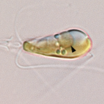
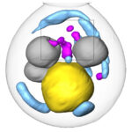
After years of work, an international team found evidence that a once-independent nitrogen-fixing microbe has become a permanent resident within algae cells.
A New Pathway for Clearing Misfolded Proteins
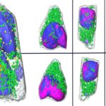
Stanford researchers have used cryogenic 3D imaging at the National Center for X-ray Tomography (NCXT) to identify a new pathway for clearing misfolded proteins from cells. This work presents a potential therapy target for age-related disorders like Alzheimer, Parkinson, and Huntington Diseases.
Molecular Biophysics and Integrated Bioimaging Division Leadership Changes
Molecular Biophysics and Integrated Bioimaging (MBIB) interim Division Director Junko Yano announced a change in leadership of the Cellular and Tissue Imaging (CTI) Department, effective January 1, 2023. Carolyn Larabell is stepping down as the Department Head, a position she has served in since July 2020. She will continue to be a member of the MBIB management team, serving as senior advisor to the Division Director. Suzanne Baker, a staff scientist whose research focuses on the methodology behind PET imaging and pharmacokinetic modeling applied to research in aging and dementia, will serve as the new CTI Department Head.
Congratulations to Biosciences Area Director’s Award Recipients
Each year, the Berkeley Lab Director’s Achievement Award program recognizes outstanding contributions by employees to all facets of Lab activities. Several Biosciences Area personnel are among the 2022 honorees.
Safely Studying Dangerous Infections Just Got A Lot Easier
A team of researchers led by NCXT Director Carolyn Larabell, in collaboration with scientists at Heidelberg University in Germany, used a technique called soft X-ray tomography (SXT) to quickly scan and analyze human lung cells infected with SARS-CoV-2. SXT not only significantly shortens the time frame, but provides more detail—increasing the chances of distinguishing subtle changes in the cell.
Was this page useful?


