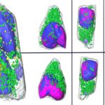
A team of researchers led by NCXT Director Carolyn Larabell, in collaboration with scientists at Heidelberg University in Germany, used a technique called soft X-ray tomography (SXT) to quickly scan and analyze human lung cells infected with SARS-CoV-2. SXT not only significantly shortens the time frame, but provides more detail—increasing the chances of distinguishing subtle changes in the cell.
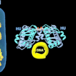
Study Gains New Insight Into Bacterial DNA Packing
A Berkeley Lab-led team of researchers used multiple high-powered X-ray techniques at the Advanced Light Source to image the process in E. coli bacteria at the micro-, meso-, and nanoscales. The imaging technique they developed enabled them to visualize the bacteria’s chromosome at higher resolutions than ever before, and without the need for labeling, which slows down the process but is required by most other techniques.
Regulation of Algal Photosynthesis and Metabolism
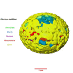
The unicellular green alga Chromochloris zofingiensis has the ability to shift metabolic modes from photoautotrophic (synthesizing food using light as energy source) to heterotrophic (obtaining food and energy from exogenous sources) in response to carbon source availability in the light. It also has the capacity—under certain conditions—to produce high amounts of commercially relevant bioproducts: notably, the ketocarotenoid astaxanthin, used in feed, cosmetics, and as a nutraceutical, and triacylglycerol (TAG) biofuel precursors.
Understanding how photosynthesis and metabolism are regulated in algae could, via bioengineering, enable scientists to reroute metabolism toward beneficial bioproducts for energy, food, and human health. To that end, Berkeley Lab Biosciences researchers used C. zofingiensis as a simple algal model system to investigate conserved eukaryotic sugar responses, as well as mechanisms of thylakoid breakdown and biogenesis in chloroplasts.
‘Minimalist’ Machine Learning Algorithms Analyze Images from Sparse Data
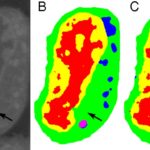
Typical machine learning methods used to analyze experimental imaging data rely on tens or hundreds of thousands of training images. But Daniël Pelt and James Sethian of Berkeley Lab’s Center for Advanced Mathematics for Energy Research Applications (CAMERA) have developed what they call a “Mixed-Scale Dense Convolution Neural Network” (MS-D) that “learns” much more quickly from a remarkably small training set. One promising application of MS-D is in understanding the internal structure and morphology of biological cells to identify, for example, differences between healthy and diseased cells. In one such project in Carolyn Larabell’s lab, the method needed data from just seven cells to determine the cell structure.
Social Media Feature Highlights 7 Lab Imaging Tools Pushing Science Forward
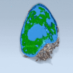
Lab scientists are developing new ways to see the unseen. Seven imaging advances, including two from the Biosciences Area, are helping to push science forward, from developing better batteries to peering inside cells to exploring the nature of the universe. The animation on the left shows a 3-D journey inside the center of cells, recently described in Cell Reports by scientists in the National Center for X-ray Tomography at the Advanced Light Source. These techniques were previously reported in the Berkeley Lab News Center and have been compiled into this listicle.
Was this page useful?


