
Amy E. Herr
Biological Faculty Engineer
Divisions
Biological Systems and Engineering
- Process Engineering & Analytics
Secondary Affiliation:
Molecular Biophysics and Integrated Bioimaging
Biography
Amy E. Herr is the Lester John & Lynne Dewar Lloyd Distinguished Professor of Bioengineering at the University of California, Berkeley and a Chan Zuckerberg (CZ) Biohub Investigator. Prof. Herr joined UC Berkeley as Assistant Professor of Bioengineering in 2007, was promoted to Associate Professor with tenure in 2012, and promoted to Full Professor in 2015. Prior to joining UC Berkeley, she was a staff member in the Biosystems Research Group at Sandia National Laboratories (Livermore, CA; 2002-2007). She earned her PhD in Mechanical Engineering at Stanford with Profs. Tom Kenny & Juan Santiago as an NSF Graduate Research Fellow, an MS in Mechanical Engineering also from Stanford, and a BS in Engineering & Applied Science from Caltech.
Professor Herr is an elected Fellow of the National Academy of Inventors and the American Institute of Medical and Biological Engineering (AIMBE), a Board Member of the Chemical & Biological Microsystems Society (CBMS) which oversees the microTAS conferences, is a standing member of the NIH Nanotechnology Study Section, and is an Advisory Board Member for the UCSF Rosenman Institute and the journals Analytical Chemistry and ACS Sensors. She has served as a Co-Director of the Cold Spring Harbor Laboratory’s Single Cell Analysis summer course (2015 & 2016), both Chair (2009) and Vice-chair (2007) of the Gordon Research Conference (GRC) on the Physics & Chemistry of Microfluidics. She is faculty advisor to the UC Berkeley chapter of the Society of Women Engineers (SWE) and the Graduate Women in Engineering (GWE).
Professor Herr’s research has been recognized by: the 2016 Mid-career Achievement Award from the American Electrophoresis Society, the 2015 Georges Guiochon Faculty Fellow from HPLC, the 2012 Young Innovator Award from Analytical Chemistry/CBMS, the 2012 Ellen Weaver Award from the Association for Women in Science (AWIS, for mentoring), a 2011 NSF CAREER award, a 2010 NIH New Innovator Award, a 2010 Alfred P. Sloan Research Fellowship in chemistry, a 2010 New Investigator Award in Analytical Chemistry from Eli Lilly & Co., a 2009 Defense Advanced Research Projects Agency (DARPA) Young Faculty Award, a 2009 Hellman Family Faculty Fund Award from UC Berkeley, a 2008 Regents’ Junior Faculty Fellowship from the University of California. Professor Herr has also been recognized by the 2012 Outstanding Instructor Award in Bioengineering (Bioengineering Honor Society student vote) and a 2007 Outstanding Mentor Award from Sandia National Laboratories.
Research Interests
Scale-dependent Phenomena Underpinning Technology Development
Large-scale study of protein structure, function, and expression (proteomics) is instrumental to molecular biomarker discovery. Due to the constantly changing nature of protein expression and state, these profiles are notoriously difficult to study. High-resolution analytical assays such as two-dimensional electrophoresis and mass spectrometry have proven essential to proteomics; nevertheless, these information-rich methods can be slow and labor intensive. With these considerations in mind, our group is developing techniques, implemented via microfluidic technologies, as a means to achieve a rapid, yet still quantitative, assessment of protein expression & state variations in complex samples.
Biomarker Validation
In spite of significant advances in proteomic technology, few new protein biomarkers have emerged from the proteomic discovery pool, progressed though the scrutiny of validation studies, and become incorporated in diagnostic tools. The long-term goal of our work is development of flexible instruments for the rapid validation of putative disease-specific biomarkers in promising diagnostic fluids. An urgent need exists for robust bioanalytical capability that delivers high-throughput validation of putative biomarkers, thus allowing subsequent incorporation of validated markers into diagnostics. To achieve this aim, our group employs nascent microfluidic technologies to seamlessly integrate complex sample preparation, sample handling, and quantitative bioanalytical assays into tools amenable to automation.
Clinical & Point-of-Care Diagnostics
Appropriate, effective biomolecular analysis mechanisms are identified for diagnostic development based upon the physicochemical characteristics of putative, disease-specific biomarkers. Most disease states are complex — diagnosis & monitoring require more than simple binary detection of a small set of proteins. To compound the difficulty in assessing disease state, analytical grade quantitation and specificity are difficult to achieve as part of a disease diagnostic, especially diagnostics employed in near-patient environments. Consequently, our group is exploring the use of electrophoretic microfluidic formats, as such formats have been demonstrated to allow rapid, analytical-grade quantitation of small sample volumes through enhanced resolving power and high-efficiency operation.
Recent Publications
Related News
Chan Zuckerberg Biohub Network Names Herr CTO
The Chan Zuckerberg Biohub Network has tapped Amy Herr, faculty engineer in the Biological and Systems Engineering Division, to serve as Chief Technology Officer. She is charged with advancing technologies to observe, measure, and analyze human biology in action.
Congratulations 2021 Chan Zuckerberg Biohub Investigators
Four faculty scientists in the Biosciences Area were included in The Chan Zuckerberg Biohub Investigator Program, awarding $21 million to 21 University of California, Berkeley researchers.
Amy Herr to Head UC Berkeley’s Bakar BioEnginuity Hub
UC Berkeley has announced a new campus initiative, the Bakar BioEnginuity Hub (BBH), that aims to launch the world-changing startups of today, while cultivating the innovative leaders of tomorrow. Opening this fall, BBH will focus on people working at the convergence of the life sciences with the physical, engineering, and data sciences. Amy Herr, a faculty engineer in the Biological Systems and Engineering (BSE) Division and UC Berkeley professor of bioengineering, has been named executive director of BBH.

Research Interests
The Holmes Lab brings techniques from machine learning, statistical linguistics, phylogenetics, and web development to bear on the interpretation and analysis of genomic data. Examples include the application of context-free grammars to understanding DNA and RNA structure; the use of phylogenetic methods in genome annotation, and to detect recombination breakpoints; the development of machine learning algorithms for bioinformatics models; the reconstruction of insertion, deletion and transposition events in genome evolutionary histories; statistical algorithms for metagenomics species distribution analysis; and dynamic-HTML web applications for collaborative genomic data analysis.
Recent Publications

Building: Advanced Light Source Building, Room 2108
Phone: 510-486-4587
Fax: 510-486-5298
JMHolton@lbl.gov
Biography
Dr. Holton is currently an Associate Adjunct Professor at the University of California San Francisco with a Faculty Associate appointment at Lawrence Berkeley National Laboratory, where he serves as the Director for the Macromolecular Crystallography (MX) X-ray Diffraction Beamline 8.3.1 at the Advanced Light Source. He is an expert in optimizing sample preparation and diffraction techniques with particular focus on radiation damage, detector performance and x-ray scattering physics. He has written several absolute-scale simulators for both “conventional” MX and femtosecond nanocrystallography that have been instrumental in designing these experiments.
Dr. Holton earned a B.S., in Biology from the California Institute of Technology and his Ph.D., in Molecular and Cell Biology from the University of California at Berkeley.
Recent Publications
Related News
Congratulations to Biosciences Area Director’s Award Recipients
Several Biosciences Area personnel are among the 2024 recipients of Berkeley Lab Director’s Achievement Awards. The program recognizes outstanding contributions by employees to all aspects of Lab activities.
Biosciences Area FY24 LDRD Projects
The projects of 21 Biosciences Area scientists and engineers received funding through the FY24 Laboratory Directed Research and Development (LDRD) program.
Researchers Capture Elusive Missing Step in Photosynthesis
After decades of effort, scientists have revealed atomic-scale details of the water splitting step of photosynthesis, the chemical process that generates the air we breathe. The latest work adds to our understanding of photosynthesis and will aid the development of fully renewable alternative energy sources.
Building: Advanced Light Source Building , Room 2136
Phone: (510) 486-5378
Fax: 510-486-5298
GLHura@lbl.gov
Research Interests
Mechanisms of biological macromolecules inspire nanoscale engineering strategies and provide insights into disease.
The speed of genomic sequencing has rapidly increased; opening up 3 billion years of evolutionary engineering, new disease treatment opportunities and providing a perspective on the complex networks involved in most biological processes. Enabled by high throughput approaches, rather than focusing on a single system or pathway I study multiple pathways and processes. I work with the view that in cellular networks there are only a few degrees of separation between the actions of any two molecules. Of particular interest are hub proteins which, through multiple interactions or adopting specific conformations, signal alternate cellular outcomes. By developing an intuition in diverse bio-macromolecular systems I also work on engineering macromolecules for new functions.
Building intuition on the large networks and multi-level feedback loops in cellular systems requires many measurements which current capabilities cannot deliver.
An understanding of mechanism has fallen behind the rate at which new molecules of interest are being identified. I utilize and develop high throughput solution based techniques to characterize the conformations biomolecules adopt in the many contexts they encounter. A primary technique has been small angle X-ray scattering or SAXS. X-rays provide access to high resolution. New light sources provide exponentially increasing power. The two aspects combined provide insights into conformations of a macromolecule in high throughput. I also develop approaches to combine crystallographic results with SAXS. Crystallography is low throughput but provides un-paralleled resolution. SAXS provides access to conformational changes in high throughput.
Publications
Related News
SIBYLS Team Recognized with Innovative Instrumentation Award
The Structurally Integrated BiologY for the Life Sciences (SIBYLS) team received the 2025 Klaus Halbach Award for Innovative Instrumentation at the 2025 Advanced Light Source (ALS) User Meeting in August.
Time-Resolved SAXS Screen of Small-molecule Drug Candidates
A team of researchers developed a high-throughput drug-discovery workflow leveraging time-resolved small-angle X-ray scattering (SAXS) capabilities at the Advanced Light Source’s (ALS) Structurally Integrated Biology for the Life Sciences (SIBYLS) beamline to identify small molecules capable of activating biomolecular dynamics associated with a desired therapeutic outcome.
University of Duisburg-Essen Delegation Explores Collaborative Opportunities with Lawrence Berkeley National Laboratory
Building on a Memorandum of Understanding signed between UDE and Berkeley Lab researchers, a kick-off meeting focused on future collaborations in the fields of genomics, structural biology, bioimaging, and water research.

Building: Life Sciences Addition, Room 271
Phone: (510) 642-9853
EYIsacoff@lbl.gov
https://vcresearch.berkeley.edu/faculty/ehud-isacoff
Links
Research Interests
Our lab’s research is focused in four intersecting and complementary areas: mechanisms of ion channel function, synapse development and plasticity, neural circuit function, and the design of novel probes for the optical detection of neuronal signaling.
Developing Tools to Study Neurons
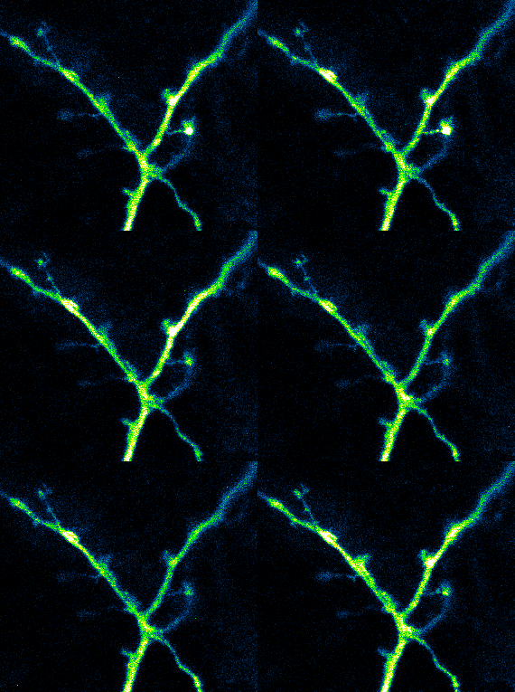
Elucidating structural and functional connectivity between neurons is one of today’s greatest challenges of systems neuroscience. We have developed a family of genetically-encoded photoactivatable calcium sensors that allow us to simultaneously visualize the morphology and activity of individual neurons, including fine structures such as dendritic spines.
Recent Publications
Related News
Biosciences Researchers Honored by the National Academy of Sciences
Three scientists affiliated with the Biosciences Area have been recognized by the National Academy of Sciences (NAS), one as the recipient of an NAS award and two as newly elected members. On Sunday, the NAS formally presented its 2018 NAS Award in Chemical Sciences to Jennifer Doudna, a faculty biochemist in the Molecular Biophysics and Integrated Bioimaging (MBIB) Division. Judith Campisi, a biochemist affiliated with Biological Systems and Engineering Division, Ehud “Udi” Isacoff, an MBIB faculty biologist, are among the group of 84 new members elected to the NAS.
DARPA Awards $21.6M to Develop Optogenetic ‘Read-Write’ Neural Interface
Ehud Isacoff of the Molecular Biophysics and Integrated Bioimaging (MBIB) Division is the project lead on a $21.6 million grant awarded to UC Berkeley as part of the Defense Advanced Research Projects Agency’s (DARPA’s) Neural Engineering System Design program. The team led by Isacoff, director of the Helen Wills Neuroscience Institute at UC Berkeley, aims to develop a novel brain-machine interface that uses light to monitor and modulate the activity of thousands to millions of individual neurons in the cerebral cortex.

Building: 91, Room 450J5
Mail Stop: 91R0183
NNIvanova@lbl.gov
https://jgi.doe.gov/who-we-are/program-platform-leads/groups/microbiome-data-science
Divisions
- Science Programs
Secondary Affiliation:
Environmental Genomics and Systems Biology
- Comparative and Functional Genomics
Recent Publications
Related News
Taking Stock of the Known and Unknown Microbial Space
In Science Advances, JGI researchers have taken stock of the current state of microbial genomic biodiversity. Using publicly available genome sequence data generated over the past three decades, their study assesses what fraction of the microbial diversity we know about, and proposes a path forward to curate and cultivate what is still unknown.
Doubling Down on Known Protein Families
Through a novel approach detailed in Nature, a massive computational analysis of microbiome datasets by the JGI focuses on unveiling protein functional diversity.
JGI Adds Actinobacteria Chapter in the Genomic Encyclopedia of Bacteria and Archaea
Large-scale comparative analysis leads to identification of biosynthetic gene clusters for novel secondary metabolites for multiple applications

Biography
Gary started his scientific career in 1978 as a research technician in the Schubiger lab at the University of Washington, where he worked on imaginal disc formation in Drosophila. He obtained his PhD in the lab of Charles Laird at the University of Washington, where he worked on the role of nucleolus organization in rDNA function. He finished his PhD in Genetics in 1987 and continued to work with Drosophila during his postdoc in the lab of Allan Spradling at the Carnegie Institute.
His work in the Spradling lab spurred his interest in heterochromatin formation and 4 years later he started his own lab at The Salk Institute, La Jolla in 1991. After being in San Diego for 12 years, during which he obtained his professorship at The Salk Institute for Biological Studies, Gary and his lab moved to the Lawrence Berkeley National Lab (LBNL) in 2003. Gary has been an adjunct professor at UC Berkeley since 2003 and became director of the LBNL Life Sciences Division in 2011.
The Karpen lab has a long-standing interest in chromatin structure and function, with a special emphasis on heterochromatic DNA regions. The current projects in the lab range from centromere formation and function, to the role of lncRNAs, ageing, and DNA repair in heterochromatin formation and maintenance.
Research Interests
Our studies are focused on understanding inheritance, chromatin structure, gene expression, and the organization of chromosomes in the nucleus. Most of our studies have focused on the fruit fly Drosophila melanogaster as a model for chromosome function in metazoans, which allows us to address mechanisms in animals by synergistically combining molecular, genetic, cell biological and biochemical approaches. Additionally, we have examined the relevance of our findings to human chromosomes, and have demonstrated surprising similarities between these evolutionarily-distant species.
Recent Publications
Related News
Gary Karpen Elected to The National Academy of Sciences
Karpen, who studies how genetic material can impact inheritance and cell function, joins the 2026 class recognized by the NAS.
Secrets of the Centromere Revealed by First Gapless Human Genome Sequence
A years-long project has illuminated the sequences at the chromosomes’ center, where proteins bind to move replicated DNA into daughter cells. The newly completed genome, dubbed T2T-CHM13, represents a major upgrade from the current reference genome, called GRCh38, which is used by doctors when searching for mutations linked to disease, as well as by scientists looking at the evolution of human genetic variation.
Exploring Human Origins in the Uncharted Territory of Our Chromosomes
A group of geneticists from Berkeley Lab, UC Davis, UC Santa Cruz, and UC Berkeley are unraveling new details about human evolution by studying the uniquely regulated portion of our chromosomes that surround the centromeres. These stretches of DNA – termed centromere-proximal regions (CPRs) – are largely composed of highly repetitive, mostly non-gene-coding sequences that […]
Research Interests
The Katz group applies emerging principles of supramolecular chemistry on solid surfaces towards the synthesis of functional advanced materials, involving catalysis and adsorption. We lead in the synthesis of organic-inorganic active sites, which are at the state-of-the-art in structural control. Our modular and generalizable engineering approach aims to roughly mimic principles of active site connectivity found in biological catalysts.
This approach leads to the highly refined design of heterogeneous catalysts, which function with an activity and selectivity that cannot be achieved using conventional approaches.
The group has most recently invented approaches for (i) mild zeolite precursor delamination, preseving framework cystallinity for applications benefiting from crystalline supports, (ii) the hydrolysis of glycosidic bonds under the mildest (i.e. close to pH 7) pH conditions that have ever been achieved, and (iii) design and synthesis of robust metal clusters that consist of an unprecedented amount of catalytic surface. Area (i) impacts catalytic applications where zeolitic supports are beneficial over traditional amorhous ones (see Grosso-Giordano et al. JACS 2018, 140, 4956—4960). Area (ii) broadly impacts the depolymerization and pretreatment of biomass to biofuels using less harsh chemicals. Area (iii) improves on the major limitation to cluster catalysts in industry—lack of stability (see Gates et al. Nature 1994, 372, 346—348).
Recent Publications

Research Interests
My research is focused on elucidating the structure and function of metalloenzymes using a combination of X-ray methods. The main emphasis is on understanding the mechanism of light-driven water oxidation in Photosystem II, a large membrane protein present in all organisms that conduct oxygenic photosynthesis. For this purpose, we use very intense femtosecond X-ray pulses as delivered by the X-ray free electron laser, Linac Coherent Light Source (LCLS), at Stanford. This technique allows us to record X-ray diffraction and X-ray spectroscopic data from the enzyme under working (physiological) conditions and provide information about the geometric and electronic structure of the catalytic site (a Mn4CaO5 cluster) of the enzyme during the water oxidation reaction. Also, we are working on generalizing these approaches to other metalloenzymes to study reaction intermediates not accessible with conventional X-ray methods.
Recent Publications
Related News
Ultrabright, Ultrashort X-ray Bursts Capture Enzymes in Action
Frame-by-frame ‘molecular movies’ reveal previously invisible intermediates, paving the way for potential new cancer treatments and antibiotics.
Mapping Photosynthesis for a Brighter Future
In this Q&A, MBIB researchers Jan Kern and Asmit Bhowmick delve into the discoveries from their recent structural biology studies of the photosynthetic apparatus in plants and bacteria.
Biosciences FY26 LDRD Projects
The Laboratory Directed Research and Development program at Berkeley Lab produces cutting-edge research for the DOE and the nation. Read about the Biosciences Area–led projects and multi-Area collaborations with Biosciences co-investigators receiving funding this cycle.

Building: 977, Room 142E
Mail Stop: 977
Phone: (510) 486-4983
Fax: (510) 486-4545
TKohwi-Shigematsu@lbl.gov
Research Interests
We have been studying a protein, SATB1, which functions as a chromatin organizer in thymocytes and activated T cells. SATB1 has a nuclear distribution resembling a cage-like structure, surrounding heterochromatin. SATB1 folds chromatin by tethering specialized genomic DNA segments, which bear an unusual physical property (base-unpairing region; BURs), to the SATB1 cage-like structure. SATB1 targets chromatin remodeling/modifying enzymes, such as HDAC1 and ISWI to BURs, and thereby regulates regional histone modification status and nucleosome positioning. Having such a function, SATB1 ablation leads to disruption of hundreds of genes, resulting in an arrest in T cell development. Our laboratory is examining the function of SATB1, its homolog SATB2, and other proteins with DNA binding specificity similar to the SATB family proteins, to see if any of them has the ability to reorganize chromatin as ES cells commit to specific cell lineages. We are interested in determining which sets of genes are targeted, how chromatin is functionally organized via looping, and how chromatin structure is marked by epigenetic modification during ES cell differentiation into the neuronal and T cell lineages.
Recent Publications

Building: 977, Room 180D
Mail Stop: 977
Phone: (510) 486-6449
Fax: (510) 486-4475
A_Kronenberg@lbl.gov
Recent Publications
Biography
Sanjay Kumar, M.D., Ph.D., was appointed Assistant Professor of Bioengineering at UC Berkeley in 2005, promoted to Associate Professor with tenure in 2011, and promoted to Full Professor in 2014. He is also Professor of Chemical and Biomolecular Engineering at UC Berkeley and a Faculty Scientist in the Biological Systems & Engineering Division of Lawrence Berkeley National Laboratory. Dr. Kumar is an elected Fellow of AIMBE and BMES. He and his research group have been fortunate to receive a number of honors, including the Presidential Early Career Award for Scientists and Engineers (PECASE), The NIH Director’s New Innovator Award, The Arnold and Mabel Beckman Young Investigator Award, the NSF CAREER Award, and the Stem Cells Young Investigator Award. Dr. Kumar has also received awards by student vote for Excellence in Graduate Advising (UCSF/UC Berkeley Joint Graduate Group in Bioengineering) and Outstanding Teaching (Bioengineering Honor Society) and has served as a Presidential Chair Teaching Fellow. Work in his laboratory has been sponsored by grants and fellowships from NIH, NSF, DOD, AHA, CRCC, LBNL, CIRM, The Beckman Foundation, the Keck Foundation, the Siebel Scholars Program, the DOE Molecular Foundry and the University of California.
Dr. Kumar earned a B.S. in chemical engineering (1996) from the University of Minnesota, where he studied lipid self-assembly in the laboratory of Matt Tirrell. He then moved on to Johns Hopkins University, where he earned an M.D. (2003) and a Ph.D. in molecular biophysics (2003) as a fellow of the NIH Medical Scientist Training Program. During the graduate portion of his training, he investigated the structure and energetics of neuronal intermediate filaments in the laboratories of Jan Hoh of the School of Medicine and Mike Paulaitis of the Department of Chemical Engineering. From 2003-2005, he served as an NIH research fellow with Don Ingber at Children’s Hospital Boston and Harvard Medical School, where he examined the nanoscale mechanics and dynamics of cytoskeletal structures in living cells and developed nanomagnetic technologies to control receptor-mediated signaling.
Research Interests
Our research team seeks to understand and control biophysical communication between cells and their surroundings. In addition to investigating fundamental aspects of this problem, we are especially interested in understanding the role played by cellular mechanobiological signaling in tumor and stem cell biology in the central nervous system.
Recent Publications
Related News
Three from Biosciences Area Named AAAS Fellows
The American Association for the Advancement of Science (AAAS), which was founded in 1848 and is the world’s largest general scientific society, announced that 489 of its members—among them nine scientists at Berkeley Lab—have been named Fellows. This lifetime honor, which follows a nomination and review process, recognizes scientists, engineers, and innovators for their distinguished achievements toward the advancement or applications of science. The three newly named Fellows from the Biosciences Area are: Sanjay Kumar, a faculty scientist in the Biological Systems and Engineering (BSE) Division; Mary Maxon, the Associate Laboratory Director for the Biosciences Area; and Len Pennacchio, a senior scientist in the Environmental Genomics and Systems Biology (EGSB) Division and the Deputy of Genomic Technologies at the DOE Joint Genome Institute (JGI).
Brown Fat Flexes its Muscle to Burn Energy—and Calories
A multidisciplinary team of bioengineers and metabolic researchers led Andreas Stahl, a professor in the Department of Nutritional Sciences & Toxicology at UC Berkeley, has figured out a new pathway that triggers brown fat to consume calories and radiate them away as heat. Sanjay Kumar, a faculty scientist in Biological Systems and Engineering (BSE) and assistant professor of bioengineering at UC Berkeley, was a co-author on the study published March 6 in Cell Metabolism. One unexpected finding was that muscle-like myosin is responsible for causing brown fat cells to stiffen in response to signals from the brain; it is this increased tension that triggers a biochemical pathway that ends with these cells burning calories for heat. “This study offers a remarkable example of how mechanical and other physical forces can influence physiology and disease in powerful, unexpected ways,” Kumar said. Understanding how brown fat is activated could unlock new ways to combat obesity. Read more from UC Berkeley News.
‘Tug of War’ Among Skin Cells Key to Development of Chicken Feathers
Sanjay Kumar, a Berkeley Lab faculty scientist in Biological Systems and Engineering (BSE), and Elena Kassianidou, a graduate student working in his lab, are co-authors on a UC Berkeley-led study published in the journal Science which for the first time linked mechanical forces acting on skin cells in a developing organism to the activation of specific genes that make the cells differentiate into more specialized types, such feathers. The researchers grew skin taken from week-old chicken eggs on artificial substrates generated by Kumar and Kassianidou to mimic the stiffness of tissues that underlie the skin in the bird. The work could pave the way to growing artificial skin for grafts that looks like normal human skin with proper spacing of hair follicles and sweat pores. Read more from the UC Berkeley News Center.


