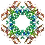
Within each cell of the human body, thousands of molecular machines are at work. They transport nutrients and biochemicals into and out of our cells, build other tiny machines, and even move our cells around. To understand how these molecular machines work, scientists create three-dimensional pictures using electron cryomicroscopy (cryo-EM), catching these machines in different shapes that give insight into their function. Now researchers at Berkeley Lab and their international collaborators who write and distribute the Phenix software suite have developed a new set of computational tools for automated structure determination from cryo-EM data.
Researchers Identify Openings for Shuttering Virus Factories
A team led by Mary Estes of the Baylor College of Medicine used rotavirus as a model to study some of the proteins involved in making the cytoplasmic compartments in which many DNA and RNA virus pathogens replicate. Banumathi Sankaran, a research scientist in the Berkeley Center for Structural Biology (BSCB) at the Advanced Light Source, collected the X-ray data at the BCSB Beamline 5.0.1 that were used to solve the three-dimensional structures of nonstructural protein NSP2. Understanding the functions of proteins that make these compartments could offer an avenue for disrupting virus production. The team published their findings in Proceedings of the National Academy of Sciences.
Programming Proteins to Pair Perfectly
Bioscientists at the Advanced Light Source (ALS) at Berkeley Lab lent their expertise to a project led by scientists at the University of Washington to design proteins in the lab that zip together like DNA. The technique could enable the design of protein nanomachines to help diagnose and treat disease, allow for more precise engineering of cells, and perform a variety of other tasks.
Toward a Blueprint for Anti-influenza Drugs
An international team led by researchers at UCSF used protein crystallography at the Advanced Light Source (ALS) beamline 8.3.1 to obtain structures of several influenza antiviral drug molecules bound to their proton-channel targets in both open and closed conformations. These complexes provide the first high-resolution views of how the drugs interact with and disrupt the water-molecule networks lining the M2 transmembrane channel. The structures provide an atomic-level blueprint from which to design more effective anti-influenza drugs that can overcome growing drug resistance. ALS beamline 8.3.1 is operated by James Holton, MBIB beamline scientist and associate adjunct professor at UCSF.
Read more in the ALS Science Highlight.
Freeze-frame Microscopy Captures Molecule’s ‘Lock-and-Load’ on DNA
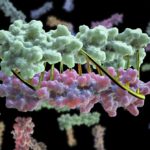
Eva Nogales, faculty scientist in Molecular Biophysics & Integrated Bioimaging (MBIB) Division and UC Berkeley professor of molecular and cell biology, led a team that captured freeze-frames of the changing shape of a huge macromolecular complex as it locks onto DNA and loads the machinery for reading the genetic code. The molecule, called transcription factor IID (TFIID), is critical to transcribing genes into messenger RNA that will later be used as blueprints to make proteins.
- « Previous Page
- 1
- …
- 35
- 36
- 37
- 38
- 39
- …
- 78
- Next Page »
Was this page useful?
Send


