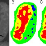
Typical machine learning methods used to analyze experimental imaging data rely on tens or hundreds of thousands of training images. But Daniël Pelt and James Sethian of Berkeley Lab’s Center for Advanced Mathematics for Energy Research Applications (CAMERA) have developed what they call a “Mixed-Scale Dense Convolution Neural Network” (MS-D) that “learns” much more quickly from a remarkably small training set. One promising application of MS-D is in understanding the internal structure and morphology of biological cells to identify, for example, differences between healthy and diseased cells. In one such project in Carolyn Larabell’s lab, the method needed data from just seven cells to determine the cell structure.
Carolyn Larabell to Receive Shirley Award at ALS User Meeting
Carolyn Larabell, a professor at the UC San Francisco School of Medicine and a faculty scientist in the Molecular Biophysics and Integrated Bioimaging (MBIB) Division at Berkeley Lab, has been selected by the Advanced Light Source (ALS) Users’ Executive Committee to receive the 2017 David A. Shirley Award for Outstanding Scientific Achievement. Larabell directs the National Center for X-Ray Tomography (NCXT) centered around ALS Beamline 2.1, the world’s first soft x-ray microscope for biological imaging, and she led the group responsible for its commissioning, design, and construction. Soft x-ray tomography makes it possible to image intact biological cells in 3-D without structure-damaging preparation such as chemically fixing, dehydrating, and staining the specimens. On October 3, Larabell will give her award talk, entitled “The Expanding Universe of Cell Biology at the NCXT,” during the ALS User Meeting at Berkeley Lab. Read more in ALS News & Updates.
3-D Imaging Technique Maps Migration of DNA-carrying Material at the Center of Cells
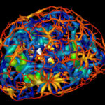
Scientists in the Molecular Biophysics and Integrated Bioimaging Division (MBIB) have produced detailed 3-D visualizations that show an unexpected connectivity in the genetic material at the center of cells, providing a new understanding of a cell’s evolving architecture. A team of researchers, led by Carolyn Larabell, MBIB faculty scientist and professor at UCSF, have mapped the reorganization of genetic material that takes place when a stem cell matures into a nerve cell using X-ray imaging tools in the National Center for X-ray Tomography at the Advanced Light Source. The results could help us understand how patterning and reorganization of DNA-containing material called chromatin in a cell’s nucleus relate to a cell’s specialized function as specific genes are activated or silenced. Read more at the Berkeley Lab News Center.
Study Finds Molecular Switch That Triggers Bacterial Pathogenicity
Scientists have revealed that the supercoiling of bacterial chromosomes around histone-like proteins can trigger the expression of genes that make the microbe invasive. The discovery could provide a new target for the development of drugs to prevent or treat bacterial infection.
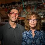
The study lead author Michal Hammel, research scientist in the Molecular Biophysics & Integrated Bioimaging (MBIB) Division, teamed up with MBIB faculty scientist Carolyn Larabell; both are pictured above. Read more on the Berkeley Lab News Center.
Was this page useful?


