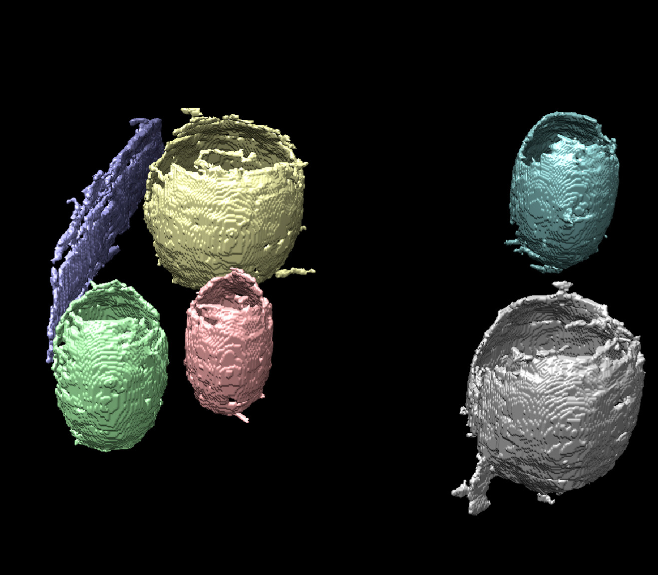
Cryo-electron tomography (cryo-ET) is a useful technology for visualizing subcellular structures at molecular resolution under near-natural conditions. Thin, flash-frozen samples are tilted at successive angles relative to an electron beam to create a series of 2D images that are then computationally reconstructed into a 3D view, or tomogram, of the sample.
However, the technique has its limitations. Contrast is inherently low due to the limited electron dose that can be applied without causing radiation damage to the sample. And because sample angles greater than about 60° would yield little useful information (due to the beam interacting with a relatively larger cross-section of the sample) they are omitted, resulting in a “missing wedge” of data.
What’s more, segmentation of these cell tomograms remains a time-consuming and challenging task—one most accurately performed by humans with extensive amounts of time on their hands. It could take one scientist several months to delineate all of the landmarks and boundaries in a tomogram and assign a label to each and every pixel.
Now, a team of Berkeley Lab scientists has developed a machine-learning pipeline to facilitate segmentation of tomograms of cell membrane structures. Given the complexity of the segmentation task and the inherent challenge in obtaining high-quality tomograms, the researchers knew that it was unlikely a single image-processing or machine-learning technique would produce satisfactory results, so they set out to develop an image analysis and segmentation pipeline that combined various methods.
The project was an LDRD-funded collaboration among Chao Yang from the Applied Mathematics and Computational Research Division, and Nick Sauter and Karen Davies from the Molecular Biophysics and Integrated Bioimaging (MBIB) Division. The team used the Cori supercomputer at the National Energy Research Scientific Computing Center (NERSC) at Berkeley Lab to test their methods and further refine the pipeline approach.
“If it were just one cell, we could do this by hand, but the real potential is to view the same cellular structure over the entire life cycle of the organism, and how it changes under different environmental conditions and external stimuli,” said Sauter. “With large data, the new machine learning techniques can help discover how the diverse ensemble of structures support function in the cell.”


