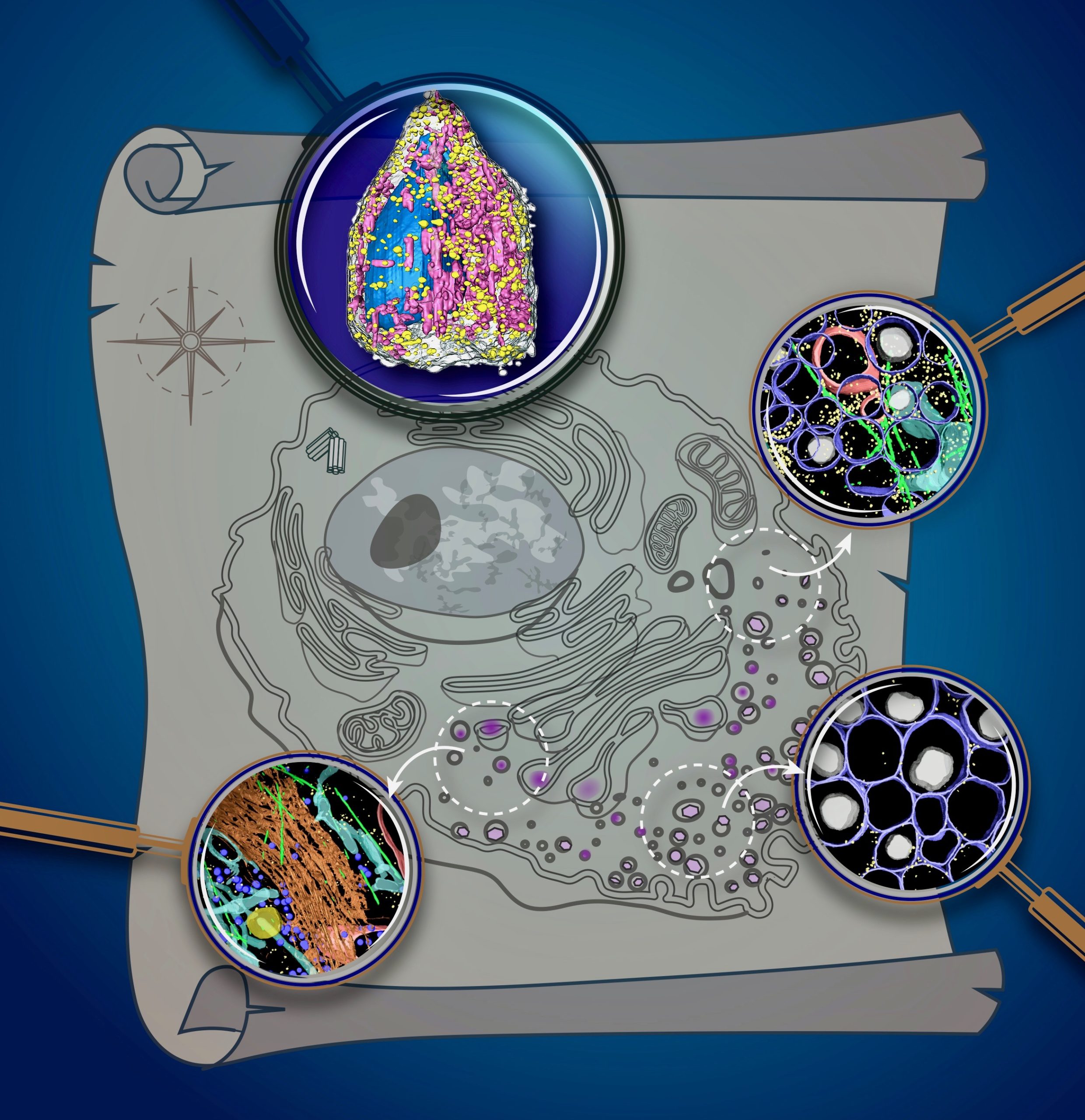
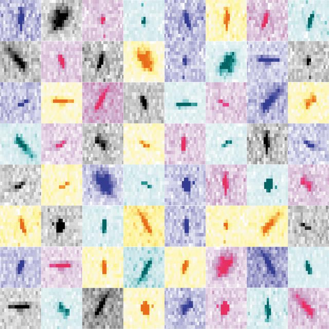
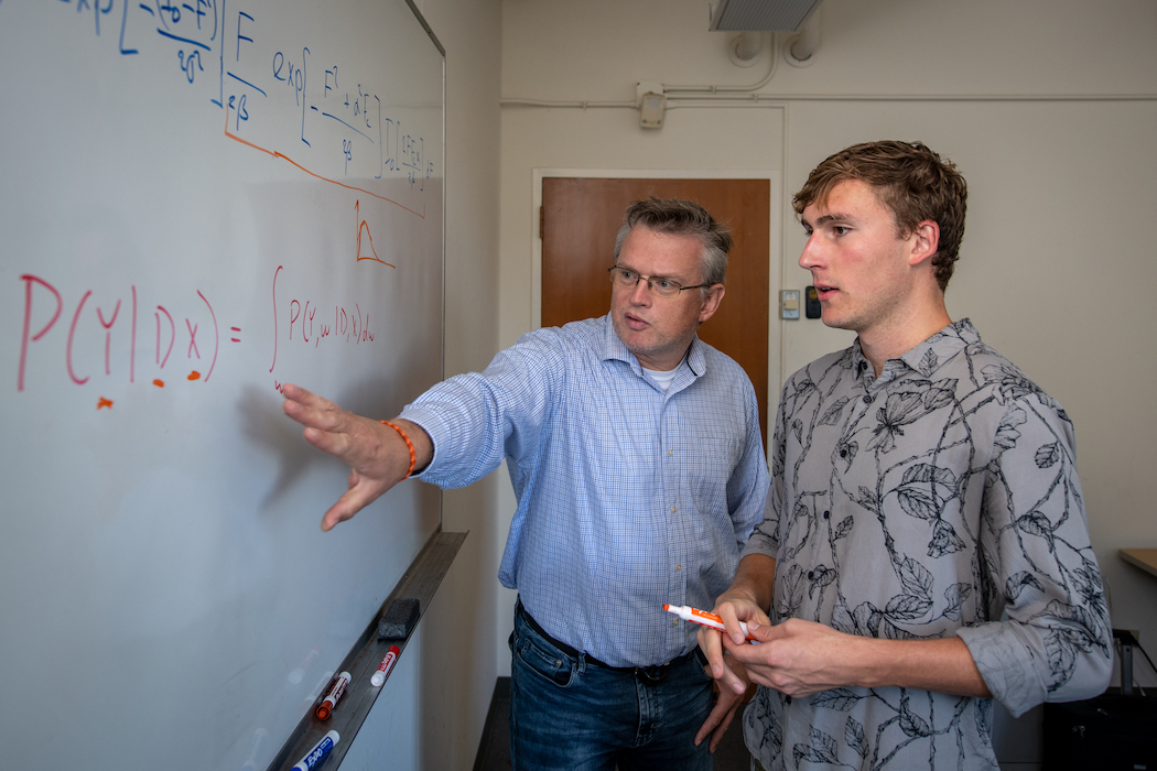
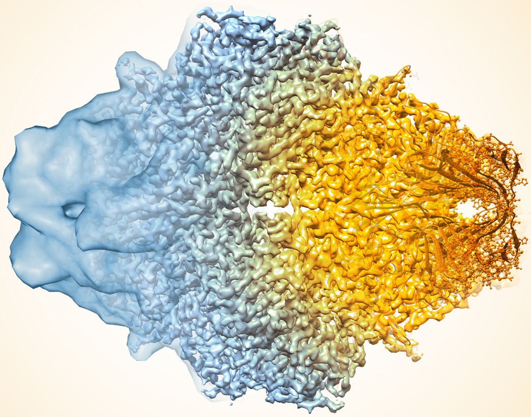
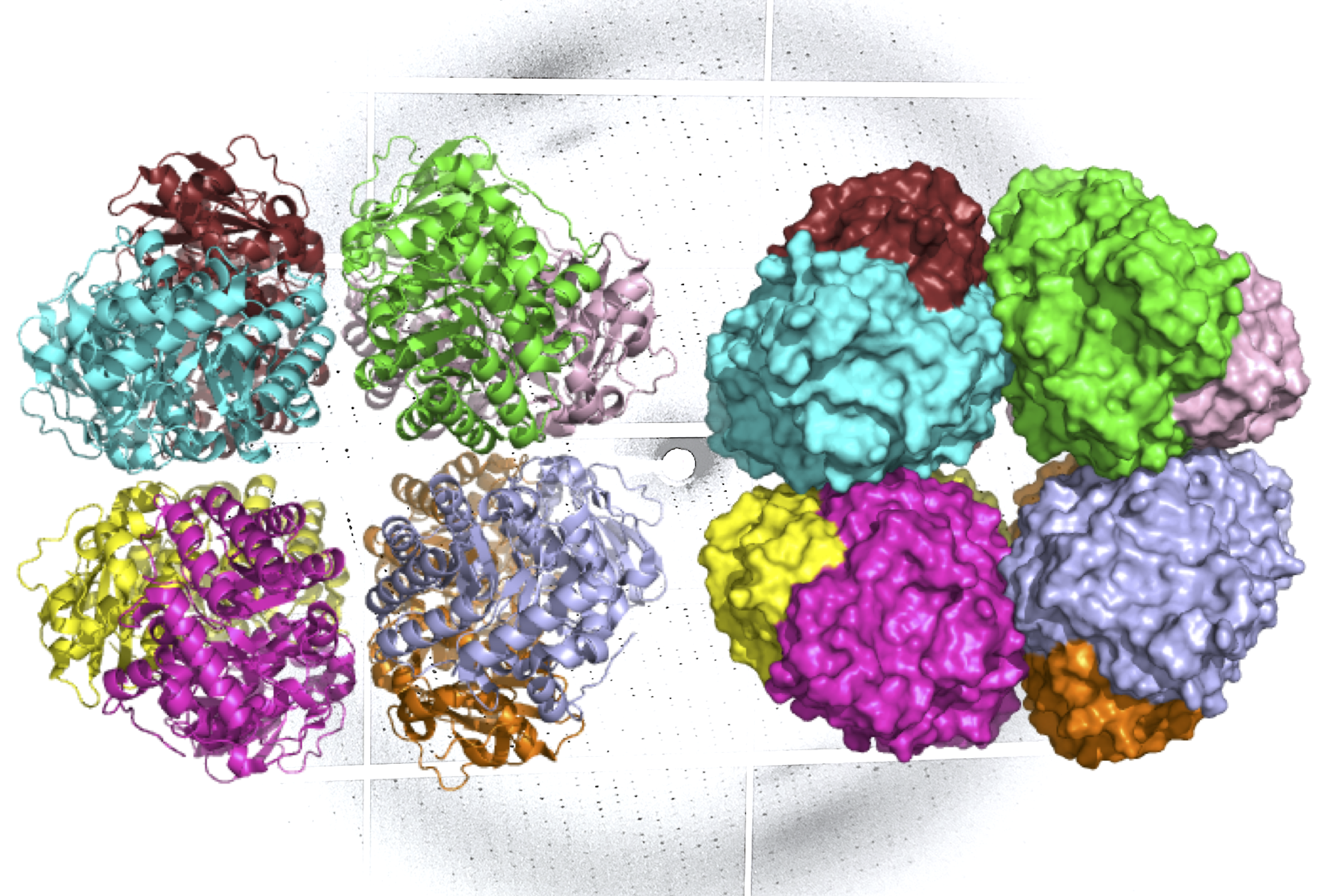
Soft X-ray tomography provides a map of organelles within an intact cell. (Credit: Katya Kadyshevskaya/USC) Collage of partial Bragg spots recorded during experiments at the XFEL. For each pixel in the `shoe-box' of a given Bragg spot, diffBragg applies precise modelling of the various parameters affecting their shape and intensity. Jennifer Doudna Univ. of Tennessee undergrad Elliot Perryman (on right) worked with Biosciences staff scientist Peter Zwart during his fall 2019 BLUR internship. (Credit: Thor Swift/Berkeley Lab) A composite image of the enzyme lactase showing how cryo-EM’s resolution has improved dramatically in recent years. Older images to the left, more recent to the right. (Credit: Veronica Falconieri/National Cancer Institute) A ribbon diagram (L) and molecular surface representation (R) of carbon-fixing form I’ rubisco, showing eight molecular subunits without the small subunits. An x-ray diffraction pattern of the enzyme, also generated by the research team, is in the background. (Credit: Henrique Pereira/Berkeley Lab) Jennifer Doudna

Was this page useful?

Send
like not like