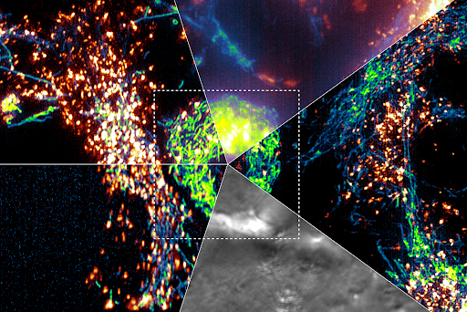
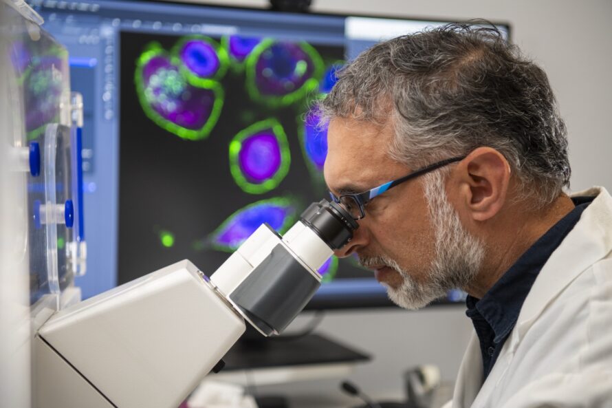
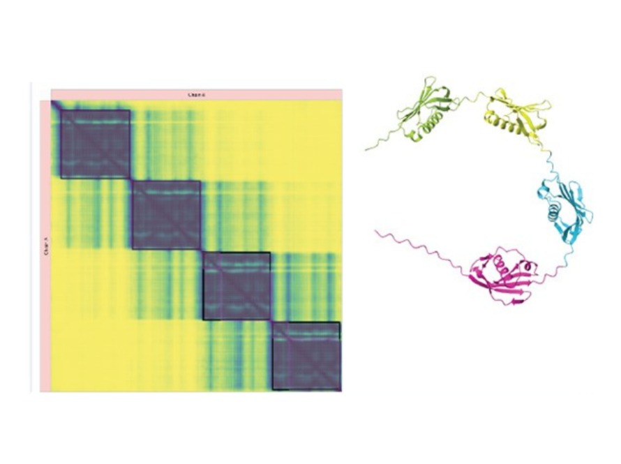
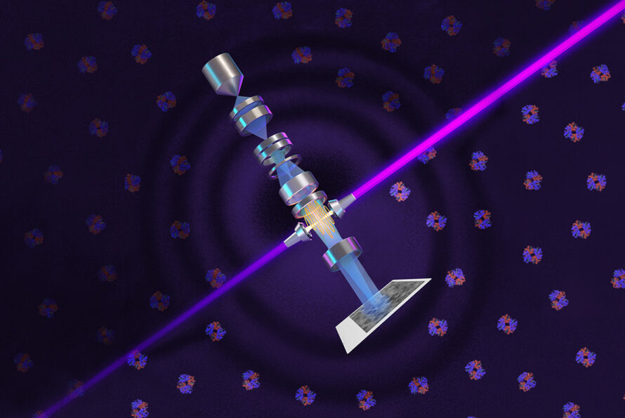
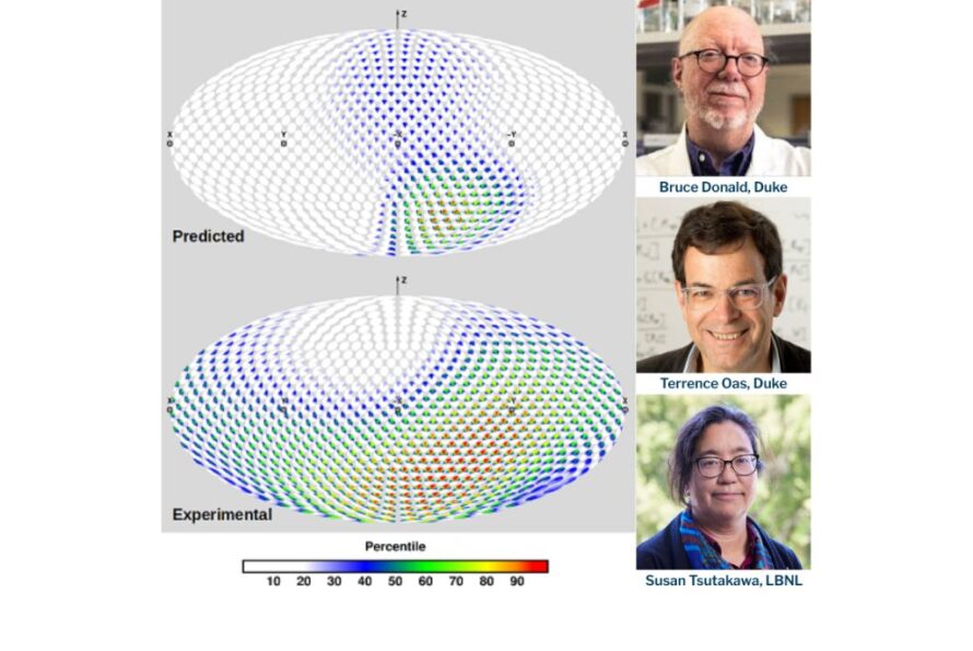
A colorful scientific composite image showing clusters of bright points and filamentous structures in five quadrants, with a central bright spot. Man looking into a microscope with multi-colored cells on a display behind him. A green frog resting on a sandy shoreline beside grass, and a bowl of oysters on ice with lemon slices next to a wooden surface. Data visualization heatmap on the left with yellow and blue gradient colors, indicating correlations, and a colorful 3D molecular structure on the right showcasing proteins. A laser (purple) is powerfully amplified by highly polished mirrors and focused on the electron beam (blue) to shift its phase and increase the cryogenic electron microscope’s contrast, allowing biologists to image smaller proteins and the crowded structures inside cells. (Credit: SayoStudio/Biohub)

Was this page useful?

Send
like not like