
Scott Classen
Computational Research Scientist

Research Interests
- Structure of Biological Macromolecules
- Beamline Control Systems
- Automation
- Artificial Intelligence and Machine Learning
Programs & Initiatives
Recent Publications
Related News
A New Angle for Studying Proteins
Researchers combined experimental data with AI and supercomputers to develop a new web-based platform that accelerates structural insights.
SIBYLS Team Recognized with Innovative Instrumentation Award
The Structurally Integrated BiologY for the Life Sciences (SIBYLS) team received the 2025 Klaus Halbach Award for Innovative Instrumentation at the 2025 Advanced Light Source (ALS) User Meeting in August.
A Fast Track for Visualizing RNA Structures
Scientists have combined multiple AI tools into a single streamlined process that predicts the atomic structures of RNA molecules.

Recent Publications
Related News
Commemorating Judy Campisi
Judith (Judy) Campisi, a leader in the field of cell senescence and a researcher at Berkeley Lab for just over 30 years, died on January 19, 2024. She was 75.
Enigmatic Protein Sculpts DNA to Repair Damage
Biosciences Area researchers and their collaborators have determined how a protein called XPG binds to and reshapes damaged DNA, illuminating its role in averting genetic disease and cancer.
Scientists Discover Protein’s Starring Role in Genome Stability, and Possibly Cancer Prevention
If you have a soft spot for unsung heroes, you'll love a DNA repair protein called XPG. Berkeley Lab scientists discovered that XPG plays a previously unknown and critical role helping to maintain genome stability in human cells. Their findings also raise the possibility that the protein helps prevent breast, ovarian, and other cancers associated with defective BRCA genes. The work, which is published online January 28 in the journal Molecular Cell, indicates XPG is essential to our health in ways far beyond it's been given credit for. Priscilla Cooper of the Biological Systems and Engineering Division conducted the research with Kelly Trego and several others at Berkeley Lab, as well as scientists from Colorado State University, Yale University, and Erasmus University Medical Center in the Netherlands. Read more at the Berkeley Lab News Center.

Divisions
Environmental Genomics and Systems Biology
- Biosystems Data Science
Secondary Affiliation:
Biological Systems and Engineering
- BioEngineering & BioMedical Sciences
Recent Publications
Related News
Foundational AI Models to Accelerate Biological Discovery
Berkeley Lab is helping build AI models for autonomous research that will enable prediction and precise design of biological systems.
Biography
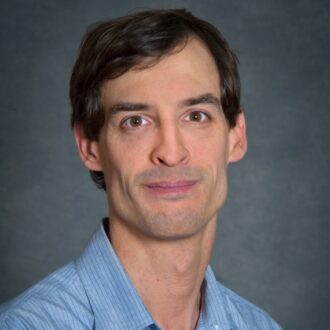
Adam Deutschbauer has a background in Microbial systems biology. As part of the Virtual Institute of Microbial Stress and Survival, he develops next-generation tools for microbial functional genomics. As the Biotechnology Component Deputy Director, he will help drive the development of experimental and computational approaches to develop models of microbial metabolism, gene regulation, and signal transduction. He will ensure the teams can meet project goals, encourage integration and collaboration between groups.
Recent Publications
Related News
EcoFABs Could Help Fuel AI in Agriculture
A first-of-its-kind global study showed that EcoFABs can deliver consistent results across labs on three continents, supported by open protocols, tools, and datasets. The reliable, large-scale data EcoFABs generate are ideal for training AI, which could help accelerate discoveries in crop development, soil health, and agriculture.
Revealing the Mysteries Within Microbial Genomes
A new technique developed by Biosciences Area researchers will make it much easier to discover the traits or activities encoded by genes of unknown function in microbes—a key step toward understanding the roles and impact of individual species.
Dub-seq Used to Screen Phage Proteins for Antibiotic Properties
A team of researchers from Berkeley Lab, UC Berkeley, and Texas A&M University worked together on a high-throughput genetic screen to identify which part of the bacteria bacteriophage viruses were targeting.

Research Interests
The Dueber Lab develops strategies for introducing designable, modular control over living cells. We are particularly interested in generating technologies for improving engineered metabolic pathway efficiency and directing flux. Our projects have applications in the development of biofuels, specialty chemicals, and environmentally friendly processes.
Recent Publications
Related News
Using Nature’s Blueprint for Sustainable Indigo Dyeing Process
Indigo has been prized since antiquity for its vibrancy and deep blue hue and, for more than a century, its unique properties have been leveraged to produce the popular textile blue denim. However, the dyeing process requires chemical steps that are environmentally damaging. A team of researchers in the Molecular Biophysics and Integrated Bioimaging (MBIB) and Biological Systems and Engineering (BSE) Divisions, at JBEI, and UC Berkeley have developed a promising sustainable indigo dyeing process that relies on genetically engineered bacteria, mimicking the natural biochemical protecting group strategy employed by the Japanese indigo plant Polygonum tinctorium.

Research Interests
Our group uses and develops advanced multidimensional ultrafast spectroscopic methods to study complex systems such as natural photosynthetic complexes, liquids, solution, and nanoscale systems such as single-walled carbon nanotubes.
In natural photosynthetic systems we aim to define the design principles underlying their remarkable .quantum efficiencies, and to use these principles to aid in the design of robust and efficient artificial photosynthetic devices. Natural systems are also regulated in response to external conditions, such as light levels, and one of the key components of Photosystem II is regularly repaired. We plan to understand the control system at the molecular level by combining molecular genetics biochemistry, modeling, and ultrafast spectroscopy through collaboration with Professor K. K. Niyogi. We have recently shown, using two-dimensional electronic spectroscopy, that long lived electronic quantum coherence exists in photosynthetic light harvesting complexes. We are exploring the implications of quantum coherence for photosynthesis and for quantum information science.
The electronic properties and excited state dynamics of nanoscale materials with significant quantum confinement effects yield a rich range of properties and potential applications. We aim to understand these properties with a particular current emphasis on single-walled carbon nanotubes via non-linear ultrafast spectroscopy and theoretical modeling.
The modern theoretical description of photochemical processes, in particular what determines which products are formed, has at its core relaxation through conical intersections. Yet very little experimental information is available on such processes. Two dimensional electronic spectroscopy has the potential to provide a window into these processes and experiments to explore conical intersection dynamics are under development.
Ultrafast multidimensional electronic spectroscopy is in its infancy with many potential ways to enhance resolution, sharpen the information content and extract specific dynamical pathways (e.g., those that involve only coherence). My group continues to develop new spectroscopic methods and the theoretical tools for their analysis.
Recent Publications
Related News
What is Quantum Biology?
MBIB's Graham Fleming recently co-authored a PNAS Perspective surveying the burgeoning field and proposing a roadmap for future research directions.
Congratulations to Biosciences Area Director’s Award Recipients
Each year, the Berkeley Lab Director’s Achievement Award program recognizes outstanding contributions by employees to all aspects of Lab activities. Several Biosciences Area personnel are among the 2025 honorees.
How Plants Manage Light: New Insights Into Nature’s Oxygen-making Machinery
A series of breakthroughs from scientists at Berkeley Lab and their collaborators provides a new understanding of how energy flows through one of nature’s most important molecular machines, the photosystem II supercomplex (PSII).
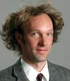
Building: 922, Room 608
Mail Stop: STANLEY
Phone: (510) 643-5624
DAFletcher@lbl.gov
http://fletchlab.berkeley.edu
Links
Research Interests
My laboratory studies the mechanics and dynamics of cell movements on the purified protein, single cell, and tissue levels. For these studies, we are developing new instruments to quantify cell and molecular mechanics based on optical microscopy, force microscopy, and microfabrication.
Recent Publications
Related News
Fletcher Elected to the National Academy of Medicine
Dan Fletcher is being recognized for the development of a mobile phone–based microscopy tool and greater contributions to our understanding of cell movement.
Congratulations 2021 Chan Zuckerberg Biohub Investigators
Four faculty scientists in the Biosciences Area were included in The Chan Zuckerberg Biohub Investigator Program, awarding $21 million to 21 University of California, Berkeley researchers.
Nitric Oxide Is the Key for Building Breast Tissue from Single Cells in 3-Dimensions
Building on four decades of research, Mina Bissell, Distinguished Scientist in Biological Systems and Engineering (BSE) Division and her colleagues have demonstrated a dynamic reciprocity between the extracellular matrix (ECM) and cell nucleus for tissue-specific gene expression. Using the 3D ECM gel to study signaling from outside the cell to the nucleus they have unraveled a dozen different pathways critical for the formation of phenotypically normal breast tissue. The signaling between the ECM and the nucleus is pivotal, bidirectional, and intricate. In two papers published in eLife this week, Bissell and Dan Fletcher, BSE faculty scientist and Purnendu Chatterjee Professor and Chair of Bioengineering at UC Berkeley, shed new light on how the extracellular matrix communicates with breast cells to generate nitric oxide, forming a loop that influences the pathway a single cell takes to form breast tissue.
Biography
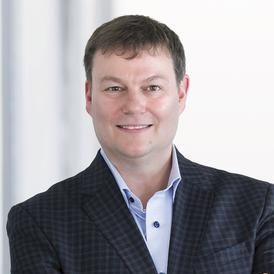
Matt Francis is the Department of Chemistry Chair and the T.Z. and Irmgard Chu Distinguished Professor in Chemistry at UC Berkeley. Matt was born in Ohio and received his undergraduate degree in Chemistry from Miami University in Oxford, OH in 1994. From 1994-1999 he attended graduate school at Harvard University, working in the lab of Prof. Eric Jacobsen. His Ph.D. research involved the development of combinatorial strategies for the discovery and optimization of new transition metal catalysts. He then moved to UC Berkeley, where he was a Postdoctoral Fellow in the Miller Institute for Basic Research in Science. He worked under the guidance of Prof. Jean Fréchet, focusing on the development of DNA-based methods for the assembly of polymeric materials and the application of dendrimers for drug delivery. Matt started his independent career in the UC Berkeley Chemistry Department in 2001, and has built a research program involving the development of new organic reactions for protein modification. These new chemical tools have then been used to modify biomolecular assemblies to prepare new materials for diagnostic imaging, wastewater treatment, and solar cell development. For his research accomplishments, Matt has received the Dreyfus Foundation New Faculty Award, an NSF Career Award, a GlaxoSmithKline Young Investigator Award, the 2017 Bioconjugate Chemistry Lectureship Award from the American Chemical Society, and the 2019 Arthur C. Cope Scholar Award from the American Chemical Society. Matt became the chair of the chemistry department at UC Berkeley in 2018. Matt has also received the UC Berkeley Departmental Teaching Award on three occasions, the Noyce Prize for Excellence in Undergraduate Teaching, and the 2009 University Distinguished Teaching Award.
Research Interests
Research in the Francis group is focused on the development of new synthetic methods for the construction of nanoscale materials. The central strategy involves the attachment of new functional components to specific locations on structural proteins, and the subsequent self-assembly of these conjugates into new types of materials with useful electronic and biological functions.
Controlled Growth of Nanocrystalline Arrays Using Cytoskeletal Proteins
Modern synthetic methods for the preparation of inorganic nanocrystals have yielded promising new components for optical and electronic device construction. However, the organization of these materials into functional assemblies remains extremely difficult, in part because the small size of nanocrystals (2-10 nm) is well below the spatial resolution of most lithographic techniques. An alternative approach could be provided by attaching these nanocrystals to specific sites on the surfaces of fiber-forming cytoskeletal proteins, such as actin. By controlling the polymerization of the actin conjugates with additional proteins and small molecule natural products, specified locations could be connected with wire-like arrays of functional materials. Once constructed, the arrays could be converted into conductive linkages, thus providing an entirely new method for nanoscale circuit construction.
Modified Viral Capsids for the Assembly of Core/Shell Materials
A second research area involves the synthesis of three-dimensional nanostructures from the self-assembling proteins that form the outer coats of viruses. For example, by selectively modifying the top and bottom faces of the satellite panicum mosaic virus capsid protein, new types of core/shell materials could be obtained after assembly. These structures could be developed into particles capable of targeting desired tissue types and releasing their cargo of drug molecules. Functionalized viral capsids could also provide new tools for the investigation of multivalent binding interactions that occur in biological systems.
New Methods for Site-Selective Protein Modification
A central theme in this research program is the modification of structural proteins in specific locations in order to achieve homogeneous and predictable assembly. Site-directed mutagenesis provides a powerful set of tools for this purpose, and will be used extensively. However, there are limitations associated with this technique, and therefore the development of new chemical approaches for protein modification will be pursued as well. This research will take advantage of the rapidly expanding set of organic reactions that can proceed in aqueous solution, and will utilize asymmetric ligands and catalysts to enhance the selectivity of protein modifications. Combinatorial reaction libraries will play an important role in this research area.
Recent Publications
Biography
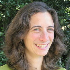
Naomi S. Ginsberg is an Associate Professor of Chemistry and Physics at University of California, Berkeley and a Faculty Scientist in the Materials Sciences and Molecular Biophysics and Integrated Imaging Divisions at Lawrence Berkeley National Laboratory, where she has been since 2010. She currently focuses on elucidating the electronic and molecular dynamics in a wide variety of soft electronic and biological materials by devising new electron and optical imaging modalities that enable characterization of fast and ultrafast processes at the nanoscale and as a function of their heterogeneities. Naomi received a B.A.Sc. degree in Engineering Science from the University of Toronto in 2000 and a Ph.D. in Physics from Harvard University in 2007, after which she held a Glenn T. Seaborg Postdoctoral Fellowship at Lawrence Berkeley National Lab. Her background in chemistry, physics, and engineering has previously led her to observe initiating events of photosynthesis that take place in a millionth billionth of a second and to slow, stop, and store light pulses in some of the coldest atom clouds on Earth. She is the Berkeley lead of STROBE, a multi-university NSF Science and Technology Center devoted to imaging science, a member of the Kavli Energy Nanoscience Institute at Berkeley, and the recipient of a David and Lucile Packard Fellowship in Science and Engineering (2011), a DARPA Young Faculty Award (2012), an Alfred P. Sloan Foundation Fellowship (2015), and a Camille Dreyfus Teacher-Scholar Award (2016) in addition to a series of teaching awards in the physical sciences. In 2017-18 she was a Miller Professor for Basic Research in Science at UC Berkeley and was designated a Kavli Fellow.
Research Interests
We are pushing the limits of spatially resolved spectroscopy and time resolved microscopy in multiple modalities, tailored to answer fundamental and challenging questions that span chemistry, physics, and biology.
- How can we investigate the optical properties of soft matter and biological systems well below the diffraction limit? Can the nanoscale dynamics of both matter and energy in these systems be studied non-invasively in real-time?
- How does the local morphology of organic electronics affect their exciton dynamics? What is the relationship between local electron dynamics and overall device performance?
- How can we learn from the remarkable efficiency of photosynthesis to guide the design and optimization of biomimetic light harvesting systems? How can energy flow be manipulated?
A common theme in our work is to investigate light-matter interactions in the near- and far-field, on ultrafast time scales, with light and electron optics, in vacuum and in the condensed phase. Please consult the Research page for additional detail.
Recent Publications
Related News
Young Biosciences Researchers Rub Elbows with Nobel Company
MBIB graduate students Margaret Doyle and Christian Tanner were among 27 highly accomplished young UC scientists selected as fellows to the prestigious 2024 Lindau Nobel Nobel Laureate Meeting in Germany where they mingled with Nobel laureates.
Naomi Ginsberg Elected Fellow of the American Physical Society
Naomi Ginsberg, a faculty scientist in the Molecular Biophysics and Integrated Bioimaging (MBIB) Division, is among the 2021 class of Fellows elected by the American Physical Society (APS). The APS Fellowship Program recognizes members who have made exceptional contributions to the physics enterprise in research, applications, leadership, service, or education. Ginsberg, who is also a UC Berkeley associate professor of chemistry, was cited for her innovative development of spatiotemporally resolved imaging and spectroscopy methods—as well as their applications in elucidating energy transport in hierarchical and heterogeneous materials and in the formation and transformation of said materials.
Tracking Energy Flow in Light-harvesting Systems on Native Nanometer and Picosecond Scales
In the first trillionths of a second after sunlight hits a photosynthetic organism, the energy that is absorbed flows through a dense network of protein-bound chlorophyll molecules to a dedicated location where it is converted to electric charges. This is the first step in a series of events that ultimately drives the formation of sugar and starch to store energy in chemical bonds. “This migration is the triggering event that leads to all of the oxygen that we breathe, all of the food that we have, and we really don’t understand why this part of photosynthesis works as well as it does. For every photon of light that’s absorbed, you can expect some biochemical action to occur. That efficiency is really remarkable,” says Naomi Ginsberg, a faculty scientist in the Molecular Biophysics and Integrated Bioimaging (MBIB) Division who has a secondary affiliation in Materials Sciences and is also a UC Berkeley associate professor of Chemistry and Physics. Ginsberg and her colleagues devised a way to measure migration efficiency, and they describe the method in Nature Materials in November 2017.

Research Interests
My lab is currently working to improve two aspects of single-particle cryo-EM where there still remains a large gap between the current state-of-the-art and what is physically achievable. The first aspect is the way in which thin specimens are spread on EM grids, prior to rapid vitrification. We are developing a number of structure-friendly ways to immobilize particles onto affinity grids, in order to prevent their adsorption at the air-water interface. At the same time, we are developing new approaches to remove as much buffer as possible while avoiding that the air-water interface touches the now-immobilized particles. The second aspect is to use an intense, focused standing wave of light as a phase plate for the electron microscope, in order to provide in-focus image contrast that is much closer to the theoretical limit than what is currently possible.
Recent Publications
Related News
Congratulations to Biosciences Area Director’s Award Recipients
Numerous Biosciences Area personnel are among the 2021 Berkeley Lab Director’s Awards honorees. This annual program recognizes outstanding contributions by employees to all facets of Lab activities. A complete list of winners can be found here. The 10th annual Director’s Awards ceremony will take place on November 18 at noon.
Congratulations to Biosciences Area Director’s Award Recipients
Numerous Biosciences Area personnel are among the 2020 Berkeley Lab Director’s Awards honorees. The award recipients are recognized for their outstanding contributions to all facets of Lab activities. The ninth annual Director’s Awards ceremony will take place (virtually) on November 12 at 3 PM.
Glaeser Honored with Glenn T. Seaborg Medal
Robert Glaeser, senior scientist in the Molecular Biophysics & Integrated Bioimaging Division, was awarded the Glenn T. Seaborg Medal by the Department of Chemistry & Biochemistry at the University of California, Los Angeles (UCLA). At a symposium held on November 1o, Glaeser and Richard Henderson, Nobel Laureate in Chemistry 2017, were recognized for their crucial contributions to the science of electron cryo-microscopy.

Biography
Jay T. Groves received his B.S. degree in Physics and Chemistry from Tufts University, and then went on to complete his Ph.D. in Biophysics with Professors Steven Boxer and Harden McConnell at Stanford University. He then spent a year as a visiting scholar at Academia Sinica in Taipei, Taiwan before becoming the Division Director’s Fellow in the Physical Biosciences Division at Lawrence Berkeley National Laboratory. In 2001 he joined the Chemistry Department at UC Berkeley as an Assistant Professor. He was promoted to Associate Professor in 2007 and Professor in 2010. In 2008 Professor Groves was appointed as a Howard Hughes Medical Institute Investigator. He has received the Burroughs Wellcome Career Award in the Biomedical Sciences (2000), the Searle Scholars Award (2002), the MIT TR100 (2003), the Beckman Young Investigator Award (2004), and the NSF CAREER Award (2005). He has served as an Associate Editor of the Annual Reviews of Physical Chemistry since 2006.
Research Interests
Professor Groves is primarily interested in role of spatial organization in biochemical reaction systems. Living cells are not at all well-mixed reaction chambers. Rather, the molecular processes of life occur in elaborate spatial patterns. This interplay between spatial organization and the chemical reactions themselves in living systems adds a fascinating new dimension to chemistry that is rarely encountered outside of biology. Specific research in Professor Groves’ laboratory focuses on how spatial organization influences signal transduction processes at the cell membrane. The research methods combine techniques in optical microscopy and spectroscopy with materials fabrication methods and cell biology. This integrated approach enables the direct observation and physical manipulation of living reaction systems, down to the single molecule level. The conceptual approach is rooted in physics and physical chemistry, with the overarching goal of developing a quantitative and mechanistic understanding biochemical processes in living systems.
Recent Publications
Related News
How T Cells Tune Out Fake Signals: Phase Transition Timing Is Everything
A group of Berkeley Lab and UC Berkeley physical chemists led by Jay Groves, faculty scientist in Molecular Biophysics and Integrated Bioimaging (MBIB), has—for the first time—imaged the process by which an individual immune system molecule is switched on in response to a signal from the environment. This breakthrough led to the discovery that the immune system activation process involves hundreds of proteins suddenly coming together to form a linked network through a process known as phase transition. Critically, the process has a built in time delay which allows the cell to distinguish a genuine receptor stimulation from background chemical noise. The work is described in a paper recently published in the journal Science.

Building: Stanley Hall, Room 274D
Phone: (510) 495-2116
OHallatschek@lbl.gov
http://hallatscheklab.berkeley.edu/
Links
Research Interests
We are trying to understand how collective patterns of self-organization emerge from the joint actions of heterogeneous individuals. The phenomena studied include evolutionary adaptation, random genetic drift, epidemic spreading, collective motion, synchronization and jamming. Although these phenomena occur in many complex systems, our experimental efforts are focused primarily on microbial systems that we can study in our wet lab. Our key theoretical challenge is to identify essential dynamical building blocks and to predict how these conspire to generate the complex dynamical patterns observed at the population level. See publications for a detailed overview of our research.


