In two recent papers, a team of scientists led by Jonathan Zehr of UC Santa Cruz (UCSC) describe the first known nitrogen-fixing organelle within a eukaryotic cell. The organelle, which they dubbed nitroplast, is the fourth example in the history of life on this planet of a prokaryotic cell being engulfed by a eukaryotic cell and evolving beyond symbiosis into an organelle.
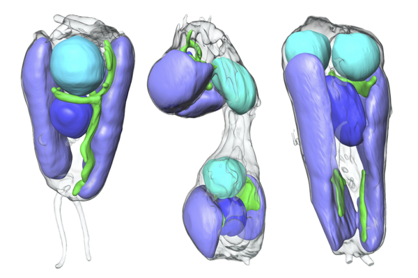
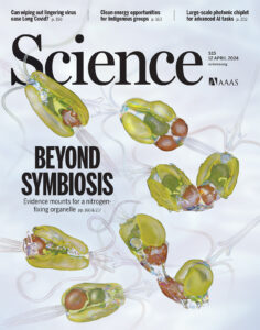
Molecular Biophysics and Integrated Bioimaging (MBIB) Division researchers, including senior faculty scientist Carolyn Larabell and research scientist Valentina Laconte, contributed to the second paper of the pair, which made the cover of the journal Science. The Biosciences Area contingent performed soft X-ray tomography (SXT) imaging on cells of the host organism, Braarudosphaera bigelowii, a marine haptophyte algae.
The technique allows scientists to rapidly visualize internal components of cells in real-time, under real-life conditions. Loconte performed the tomography, then analyzed the data to generate detailed images showing the prospective organelle’s movements within the alga at all stages of replication.

“We showed that the process of replication and division of the algal host and endosymbiont is synchronized,” said Larabell, Director of the National Center for X-Ray Tomography, who co-developed the advanced soft X-ray tomography approach at Berkeley Lab’s Advanced Light Source (ALS).
A follow on to a study published in Cell last month that demonstrated linkage of host and endosymbiont metabolisms, the Science paper also showed that the organelle-like structure relies on proteins from the host, work performed by UCSC postdoctoral scholar Tyler Coale. Taken together, the evidence led the team to confidently conclude that the erstwhile endosymbiont is, in fact, a bona fide organelle.


