Carlos Bustamante
Biophysicist Faculty Scientist

Building: 922, Room 608A
Mail Stop: MC#3220
Phone: (510) 643-9706
Fax: (510) 643-4500
CJBustamante@lbl.gov
Links
Research Interests
Our lab specializes in single molecule biophysics, where we can track and measure the activity of individual enzymes. By looking at each enzyme, we can parse out effects which are not resolvable in bulk experiments. We use optical tweezers, magnetic tweezers, atomic force microscopy (AFM), single molecule fluorescence, fluorescence correlation spectroscopy (FCS), and super-resolution photo-activatable light microscopy (PALM).
We are interested in studying how the cell converts chemical energy into mechanical work through highly specialized molecular machines. The generation, transduction, and regulation of force are key to many central processes in the cell. Many enzymes, such as polymerases, are motors which move along a cellular track, using chemical energy to take regulated steps and control synthesis.
Recent Publications
Related News
Carlos Bustamante Wins Biophysical Society’s 2021 Kazuhiko Kinosita Award
Carlos Bustamante, a biophysicist faculty scientist in the Molecular Biophysics and Integrated Bioimaging (MBIB) Division, received the 2021 Kazuhiko Kinosita Award during the Biophysical Society's 65th Annual Meeting in February.
Building: 977, Room 104
Mail Stop: 977
Phone: (510) 486-7273
MRStampfer@lbl.gov
http://hmec.lbl.gov
Links
Research Interests
The long-term objective of my program since 1976 has been to develop and characterize an experimentally tractable human mammary epithelial cell (HMEC) culture system for use in a wide variety of studies on normal human cell biology, aging, and carcinogenesis. Our aim has been to understand normal HMEC processes, and how these processes may be altered during immortal and malignant transformation, and during aging. We believe that understanding normal healthy biology is necessary for clear understanding of what constitutes abnormal processes. Our desire to facilitate widespread use of human epithelial cells for molecular and cellular biology studies has led us to develop an HMEC system that is relatively easy to use, can provide large quantities of standardized cell populations, and is well-characterized. Ongoing collaborative studies assessing the effects of cell-cell and cell-ECM components aim to improve the ability of this in vitro culture system to accurately reflect in vivo biology.
To address our goals, we have generated an integrated HMEC culture system containing:
- Normal finite lifespan HMEC
a) The normal HMEC display long-term growth, ~30-60 population doublings
b) Cells are available from women aged 16-91, allowing study of aging effects.
c) Multiple lineages (myoepithelial, luminal, progenitor) are present, allowing study of lineage differentiation. - Aberrant finite lifespan HMEC
a) Normal HMEC were exposed to a variety of oncogenic agents/genomic changes reflective of known in vivo breast cancer etiology (e.g., chemical carcinogens, stress, p53 or p16 inactivation).
b) Exposed cultures produced aberrant cells that bypassed or overcame a stress-associated senescence barrier (stasis) via errors in the RB pathway, to become aberrant post-stasis cultures.
c) A variety of distinct post-stasis phenotypes were generated, reflecting different potential types of in vivo pathways of malignant progression. - Immortally transformed lines isogenic to normal and aberrant finite HMEC
a) Post-stasis cultures produced rare clonal lines possessing genomic errors and a variety of distinct phenotypes, including both basal and luminal.
b) Non-clonal lines without gross genomic errors have been derived by direct targeting of tumor-suppressor barriers (stasis, replicative senescence).
c) Since non-malignant immortally transformed lines have overcome the major tumor suppressor barriers, including oncogene-induced senescence (OIS), they can be readily transformed to malignancy following exposure to specific oncogenes.
d) We also have hTERT-immortalized lines, however they display very long telomeres and high telomerase activity, unlike any normal HMEC or HMEC with cancer-associated immortalization (short-regulated telomeres, modest telomerase activity); we do not think they are a good model for either normal or cancer.
Detailed information on the derivation, characterization, and methods for growth of these cells, as well as information on how other labs may obtain these cells, can be found on my website: http://hmec.lbl.gov
Our laboratory’s long-term emphasis on extensive development and characterization of one human epithelial cell type model system has enabled a unique overview and led us to produce a new model of the tumor-suppressive senescence barriers encountered by cultured normal finite lifespan HMEC as they grow, senesce, overcome senescence barriers, and gain immortality and malignancy. These ongoing studies have indicated that most (not all) normal cultured HMEC cease proliferation due to a stress-associated senescence barrier, stasis, mediated by the retinoblastoma protein (not telomere attrition). Normal HMEC prior to this barrier (pre-stasis) display significant biological differences compared to finite post-stasis HMEC or to isogenic fibroblasts.
Immortality derives from overcoming the replicative senescence barrier resulting from telomere attrition, and requires reactivation of endogenous telomerase activity – which also confers resistance to OIS. Both pre- and post-stasis finite HMEC show many significant differences compared to non-malignant immortally transformed lines, whose molecular phenotype more closely resembles cancer-derived lines than finite cultures. Significantly, our in vitro transformation model is consistent with the molecular changes observed during malignant transformation in vivo; e.g.; the molecular and genomic properties of cells in hi-grade DCIS, where immortalization is first observed, closely resemble cognate cancer, more than normal cells. Our model system makes possible examination of factors that may enhance or inhibit carcinogenesis at different stages of progression.
Recent Publications
Related News
Normal is Good: Breast Cells Produced in Novel Media Resemble Those In Vivo
When studying human cells in a laboratory, it is important that the media, or the broth that bathes the cells, contains all of the nutrients necessary to support cells through their normal growth and division phases even though they are outside of the body. Bioscientists at Berkeley Lab have a long history of studying breast cancer, and Martha Stampfer, senior scientist in the Biological Systems & Engineering (BSE) Division, has spent decades developing media now widely used by the community. Today, PLOS ONE published a study describing a comprehensive analysis of three kinds of media used to grow human mammary epithelial cells (HMEC).
Building: 1, Room 360
Mail Stop: DONNER
Phone: (510) 486-7469
Fax: (510) 486-6488
PJWalian@lbl.gov
Links
Research Interests
Our group maintains a long-standing interest in the structure and function of macromolecular complexes, with an emphasis on membrane protein biology. Areas of investigation have included molecular mechanisms facilitating solute transport, intra-membrane proteolysis, cell-cell interactions, and protein-protein interaction networks. As a member of the ENIGMA SFA program, a significant portion of our research has focused on the identification and characterization of cell membrane-based mechanisms utilized in bacterial stress response, and the formation and maintenance of microbial communities. The subjects of these studies have been characterized using a range of biophysical and biochemical approaches involving methods such as the large-scale purification of recombinant and endogenously expressed proteins, liposome reconstitution, electron microscopy, X-ray and electron crystallography, X-ray micro-computed tomography (micro-CT), molecular dynamics simulations and proteomics.
Recent Publications

Building: 91, Room 110H2
Mail Stop: 100PGF100
Phone: (480) 225-2341
BPBowen@lbl.gov
http://openmsi.nersc.gov
Links
Research Interests
Understanding biological mechanisms associated with change.
Understanding complex, dynamic metabolic networks in an environmental context will require utilization of emerging technologies. These datasets generated are often large-scale both in terms of complexity and raw-size making them difficult to mine for biological insight. Ben is leading the OpenMSI and Metabolite Atlas efforts to make the most high-performance, advanced data management, model building, analysis and visualization resources for mass spectrometry accessible to all scientists via the web.
Programs & Initiatives
- OpenMSI
- Metabolite Atlas
- Biochemical Modeling
- Compound Discovery and Identification
Recent Publications
Related News
EcoFABs Could Help Fuel AI in Agriculture
A first-of-its-kind global study showed that EcoFABs can deliver consistent results across labs on three continents, supported by open protocols, tools, and datasets. The reliable, large-scale data EcoFABs generate are ideal for training AI, which could help accelerate discoveries in crop development, soil health, and agriculture.
When Marine Algae Get Sick: How Viruses Shape Microbe Interactions
Researchers in the Environmental Genomics and Systems Biology Division collaborated on a study to better understand the role of viruses that infect photosynthetic phytoplankton in the marine food web.
EcoFAB: A Tool for Combating Climate Change and Training the Next Generation
Fabricated ecosystems—EcoFABs—are plastic, takeout box–sized growth chambers developed at Berkeley Lab to be a standardized and reproducible platform for conducting experiments on model plants and the microbes that live around their roots. A greater understanding of how plants and microbes work together to store vast amounts of atmospheric carbon in the soil will help in the design of better bioenergy crops for the fight against climate change.
Building: 64, Room 242
Phone: (517) 432-4371
CKerfeld@lbl.gov
http://www.kerfeldlab.org
https://en.wikipedia.org/wiki/Cheryl_Kerfeld
Links
Divisions
Environmental Genomics and Systems Biology
- Comparative and Functional Genomics
Secondary Affiliation:
Molecular Biophysics and Integrated Bioimaging
- Structural Biology
Recent Publications
Related News
Toward Engineering Cell-like Factories
Cheryl Kerfeld’s laboratory, which operates at both Michigan State University and Berkeley Lab, teamed up with researchers at Pennsylvania State University and the University of Delaware to take a first step toward creating artificial cells that lack a lipid membrane.
Kerfeld to Lead New DOE-funded Center for Catalysis in Biomimetic Confinement
With $10.65 million in support from the U.S. Department of Energy (DOE), a new Energy Frontier Research Center based at Michigan State University (MSU) has been established. Led by Cheryl Kerfeld, a professor in the MSU-DOE Plant Research Laboratory, the Center for Catalysis in Biomimetic Confinement (CCBC) will explore how nature compartmentalizes some of its most important biochemical reactions.
Cryo-EM Reveals Blueprint for Microbial Photosynthesis
Advances in cryogenic electron microscopy have enabled an international team of experts to visualize the structure of a cyanobacterial phycobilisome with nearly atomic resolution. The work, a collaboration among researchers at Michigan State University, UC Berkeley, Berkeley Lab, and the University of South Bohemia in the Czech Republic, was published in Nature. Knowing the position of different proteins and pigments helps scientist better understand this natural process and can inspire future applications in areas such as renewable energy and environmental remediation.
Building: 6, Room 2120
Mail Stop: 6R2100
Phone: (510) 486-6511
Fax: (510) 486-5664
BSankaran@lbl.gov
Links
Research Interests
I am in charge of the the Collaborative Crystallography (CC) program at the Advanced Light Source (ALS). The program aids for a fast, reliable and transparent mail-in crystallographic service for the structural biology community. One of the key features of the CC program is that all the service work is peer reviewed by the scientific community via the ALS general users proposal system. The main benefit of having the service work peer reviewed, is that it allows for a prioritization of proposals on the basis of scientific excellence. The nature of the service work we perform differs from project to project, but can involve all steps from data collection all the way up to validation and submission of the structure to the PDB. Recently, we have been trying to expand our service to include crystallization as well. Users that participate in the CC program agree to include the ALS scientist involved as a coauthor on related manuscripts and on the PDB deposition (more than 200 to date). This program yielded 67 CC-related publications with a total citation count of just over 550, illustrating the productivity and relevance of the program.
Over the last two years, I have worked with more then 20 user groups within the United States. The goal of the CC program is to provide further support to the biological and life sciences community, in particular to those researchers who can benefit from structural information but do not have routine access to equipment and expertise needed to go from pure protein (or gene) to 3D structure. We are currently working on proof-of-principle case studies highlighting the strengths of these expanded services and will pursue opportunities to obtain sustained external funding to continue this effort.
Recent Publications
Related News
A Promising Compound for Reversible Male Contraception
Researchers at the Baylor College of Medicine identified a small-molecule protein inhibitor that blocks an enzyme that is key to male fertility. Protein crystallography performed at the Advanced Light Source (ALS) beamline 5.0.2 provided valuable structure-activity insights.
Researchers Gain Mechanistic Insight into a Viral-factory Protein
A team that included Banumathi Sankaran of the Molecular Biophysics and Integrated Bioimaging Division studied a protein called σNS, an important component of some viral factories. Understanding how this protein works will foster development of therapeutic strategies against viruses that use similar proteins to replicate.
BCSB Confirms Design of Stimulus-responsive, Two-state Proteins
AI-generated hinge proteins could open the door to solving complex challenges in the world of protein design.

Divisions
- Science Programs
Secondary Affiliation:
Environmental Genomics and Systems Biology
- Comparative and Functional Genomics
Research Interests
Genomics of fungi and algae, computational biology
Recent Publications
Related News
Oil-Rich Alga Reveals Ideal Platform for Bioengineering
Streamlined genome with precise gene targeting transforms oil-producing alga into a powerful platform for developing bio-based industrial products.
Mapping the Earth’s Hidden Fungal Kingdom
The JGI is leveraging massive comparative genomics to decode millions of mystery genes and lay a future-ready foundation.
Ancient Algae Reveal Secrets of Plant Evolution
Research uncovers the genetic foundations that helped plants conquer land over 600 million years ago.
Building: Stanley Hall, Room 708C
Mail Stop: STANLEY
Phone: (510) 642-0557
Fax: (510) 666-3336
ENogales@lbl.gov
Links
Biography
Eva Nogales is a senior faculty scientist, Professor of Biochemistry, Biophysics and Structural Biology, and a Howard Hughes Investigator. The Nogales Lab is dedicated to gaining mechanistic insight into crucial molecular processes in the life of the eukaryotic cell. Their two main research themes are the dynamic self-assembly of cytoskeleton during its essential functions in cell division, and the molecular machines governing the regulation of gene expression, specially at the transcriptional level. The unifying principle in their work is the emphasis on studying macromolecular assemblies as whole units of molecular function by direct visualization of their architecture, functional states and regulatory interactions. With this overall aim in mind they use electron microscopy and image analysis, complemented with biochemical and biophysical assays, towards a molecular understanding of their systems of interest.
Research Interests
cryo-EM, biochemistry, complex biological assemblies, structure and regulation of the cytoskeleton, microtubule dynamics, human transcriptional initiation machinery, epigenetics, gene silencing, biophysics
Recent Publications
Related News
Nogales Elected to UK’s Royal Society
Biophysicist elected to one of the world’s oldest scientific academies.
Nogales Garners Two International Accolades
In December, Eva Nogales, senior faculty scientist Molecular Biophysics and Integrated Bioimaging (MBIB) Division, traveled to her native Spain where she was honored for her work in the field of visualization of macromolecular function.
A Better Understanding of DNA Unpacking
Using specialized equipment, including cryo-EM researchers were able to study the atomic structure of a complex that allows enzymes to access DNA. A deeper knowledge of how it works could help develop better therapies for cancer and Alzheimer’s.
Research Interests
Pereira’s research focuses on protein X-ray crystallography to understand at the atomic level the function of a diverse range of enzymes related to several Joint BioEnergy Institute projects from Deconstruction, Feedstocks, Biofuels and Bioproducts, and Technology Divisions.
Recent Publications
Related News
Newly Discovered Bacterial Enzyme Produces Useful Biopolymer
Biosciences researchers collaborated on a University of British Columbia-led study that identified a bacterial enzyme that produces a novel biopolymer which holds promise as a useful biomaterial because of its biodegradability and biocompatibility.
AI-Fueled Software Reveals Protein Structure Prediction
Structural biologists have applied powerful machine learning methods to a library of protein structures to predict a protein’s shape from its gene sequence.
Study Finds ‘Missing Link’ in the Evolutionary History of Carbon-Fixing Protein Rubisco
In a study appearing in Nature Plants, researchers from UC Davis, UC Berkeley, and Berkeley Lab report the discovery and characterization of a previously undescribed lineage of form I rubisco – one that the researchers suspect diverged from form I rubisco prior to the evolution of cyanobacteria. The novel lineage, called form I’ rubisco, gives researchers new insights into the structural evolution of form I rubisco, potentially providing clues as to how this enzyme changed the planet. The work was led by Patrick Shih, a UC Davis assistant professor and the director of Plant Biosystems Design at the Joint BioEnergy Institute (JBEI), and Doug Banda, a postdoctoral scholar in his lab.

Building: 978, Room 4256
Mail Stop: 978-4121
Phone: (510) 486-7237
CJPetzold@lbl.gov
https://www.jbei.org/person/chris-petzold/
Links
Biography
Chris Petzold is a staff scientist within Berkeley Lab’s Biological Systems and Engineering Division. He earned his Ph.D. in Chemistry from Purdue University, specializing in gas-phase ion chemistry and mass spectrometric methods development. Following his doctoral studies, he conducted post-doctoral research at UC-Berkeley, applying mass spectrometry to the fields of glycomics and lipidomics. In 2005, Chris joined Jay Keasling’s research group to lead efforts in understanding the systemic impacts of metabolic engineering on host microbes.
As the Director of the Functional Genomics Research Group at the Joint BioEnergy Institute (JBEI), Chris focuses on developing mass spectrometric solutions to the complex challenges of biofuel and bioproduct manufacturing. His expertise lies in bridging high-throughput analytical technologies with advanced metabolic engineering to enable the rational design and optimization of novel biosynthetic pathways.
Research Interests
Bioanalytical Mass Spectrometry, Proteomics, Analytical Chemistry, Metabolic Engineering
Current Projects & Expertise
- Automated DBTL Workflows: Integrating laboratory automation for reproducible data acquisition to enable machine learning (ML) and Artificial Intelligence to rapidly navigate vast parametric design spaces. Recent work includes guiding the development of automated methods for microbial culture growth, sampling, data acquisition, and analysis.
- High-Throughput Screening (HTS) & RapidFire-MS: Implementing high-throughput mass spectrometry (RapidFire-MS) for the HTS of microbial libraries. This includes quantifying diverse targets such as isoprenol, isoprenol acetate, 3-hydroxypropionic acid (3-HP), and various other bioproducts to accelerate biomanufacturing timelines.
- Advanced Proteomics Methods: Implementing high-throughput targeted and shotgun proteomics to validate gene perturbations (e.g., CRISPRi effectiveness) and uncover biological mechanisms driving production improvements.
- Data Management & Interoperability: Leading JBEI efforts in biological data management to enable AI/ML workflows. This includes developing robust systems for automated data import, ensuring data interoperability through metadata capture and strong ontologies, and fostering data sharing.
Recent Publications
Related News
Biological Systems and Engineering Division and Program Leadership Changes
Division Director Blake Simmons announced that, effective March 2, Chris Petzold will lead the Biodesign Department as Interim Head following Nathan Hillson’s departure. Petzold will also assume the role of Chief Information Officer for the Joint BioEnergy Institute, as announced by CEO Jay Keasling. Katy Christiansen will serve as the lead principal investigator of the Agile BioFoundry.
Foundational AI Models to Accelerate Biological Discovery
Berkeley Lab is helping build AI models for autonomous research that will enable prediction and precise design of biological systems.
ABF, ABPDU Scale up Acrylic Acid Precursor with Industrial Microbes
Researchers at the Agile BioFoundry (ABF) and the Advanced Biofuels and Bioproducts Process Development Unit (ABPDU), have successfully scaled up a bio-based process that converts ethanol into a valuable precursor for consumer products, such as paints, coatings, and diapers. The researchers worked with industry partner, Industrial Microbes, to develop the process.
Research Interests
Miaw-Sheue Tsai is the Director of the Expression and Molecular Biology (EMB) Core for the SBDR (Structural Cell Biology of DNA Repair Machines) Program Project. SBDR brings together the top laboratories in DNA repair through multi-disciplinary collaborations to define the complex and dynamic network of DNA repair machines and to develop an actionable, quantitative and mechanistic knowledge of DNA repair machines, pathways and intersections with DNA replication and other DNA related processes to aid prediction and intervention for cancer biology. The EMB Core has been an integral part of SBDR since 2001 and is designed to lower the barriers for SBDR to obtain challenging DNA repair proteins and complexes by providing centralized resources and expertise in advanced recombinant protein expression technologies. The research and production services of the EMB Core deliver consistent, high quality key reagents (including vectors, proteins and cell products) and protein partnership information to jump-start the proposed work by the SBDR Program and facilitate both internal and external collaborations.
Recent Publications
Related News
Miaw-Sheue Tsai, DNA Mender
A research scientist in the Biological Systems and Engineering Division, Miaw-Sheue Tsai leads a group that focuses on learning how cells identify and correct damaged DNA. Originally from Taiwan, Tsai worked alongside an encouraging supervisor and discovered a place for herself in science.
Congratulations to Biosciences Area Director’s Award Recipients
Several Biosciences Area personnel are among the 2019 Berkeley Lab Director’s Awards honorees. This annual program recognizes outstanding contributions by employees to all facets of Lab activities. A complete list of winners can be found here. The Director’s Achievement Awards ceremony will take place on November 15 at 4 PM in the Building 50 Auditorium. All staff are invited and the event will be streamed live.
Strong Biosciences Presence at East Bay STEM Career Awareness Day
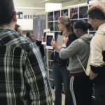 The 7th Annual East Bay STEM Career Awareness Day took place on April 26 at Wareham Development’s Aquatic Park Center in West Berkeley, home to Biosciences Operations @ Berkeley and several Area research groups. The event, organized by the Institute for STEM Education housed at California State University East Bay, had support from Berkeley Lab and several East Bay-based businesses.

Building: 84, Room 240
Mail Stop: 84R0171
Phone: (925) 980-3711
Fax: (510) 486-4229
LAPennacchio@lbl.gov
Links
Divisions
- Genomic Technologies
Secondary Affiliation:
Environmental Genomics and Systems Biology
- Comparative and Functional Genomics
Research Interests
Defining the vast landscape of gene regulatory sequences in the human genome.
Understanding how variation in regulatory sequences influences human disease/biology.
Assessing and exploiting next generation sequencing technologies for applications in both the energy and health sectors.
Recent Publications
Related News
AI Helps Decode Gene Regulation
A combination of comprehensive experiments and machine learning is uncovering hidden complexities of gene expression during development.
The Paradox of ‘Ultraconserved’ Enhancers: Perfect Sequence Conservation Not Required
The last common ancestor of humans and rodents lived more than 80 million years ago, and billions of changes in their respective DNA sequences have occurred over this vast timespan. Yet, intriguingly, there are a few hundred stretches of DNA in our genome that are still identical to the corresponding sequences in mice and rats. Generally, sequence conservation between distantly related species is an indication that the function the DNA serves is vitally important and highly sensitive to mutations. For example, most DNA sequences that encode proteins show at least moderate conservation in evolution. However, more than two-thirds of the “ultraconserved” sequences shared by humans and rodents are outside of protein-coding genes, raising the question of what led to their extreme level of conservation.
Three from Biosciences Area Named AAAS Fellows
The American Association for the Advancement of Science (AAAS), which was founded in 1848 and is the world’s largest general scientific society, announced that 489 of its members—among them nine scientists at Berkeley Lab—have been named Fellows. This lifetime honor, which follows a nomination and review process, recognizes scientists, engineers, and innovators for their distinguished achievements toward the advancement or applications of science. The three newly named Fellows from the Biosciences Area are: Sanjay Kumar, a faculty scientist in the Biological Systems and Engineering (BSE) Division; Mary Maxon, the Associate Laboratory Director for the Biosciences Area; and Len Pennacchio, a senior scientist in the Environmental Genomics and Systems Biology (EGSB) Division and the Deputy of Genomic Technologies at the DOE Joint Genome Institute (JGI).

