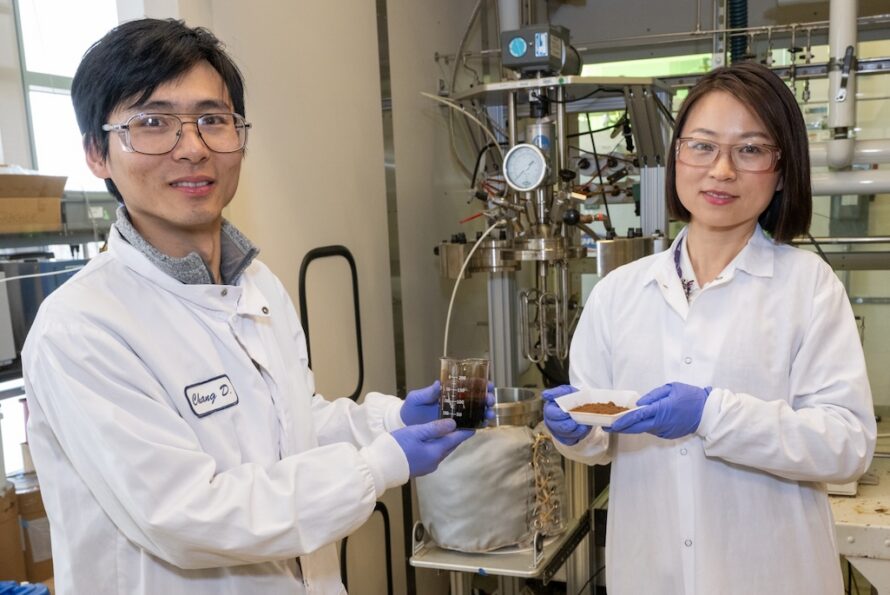
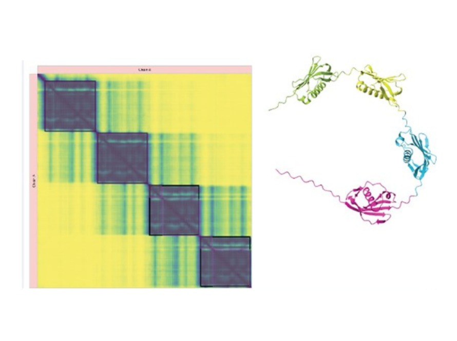
Data visualization heatmap on the left with yellow and blue gradient colors, indicating correlations, and a colorful 3D molecular structure on the right showcasing proteins. A robotic arm hovers over multiple transparent containers in a lab, holding a case with two green plant shoots, illuminated by soft green and blue light. A laser (purple) is powerfully amplified by highly polished mirrors and focused on the electron beam (blue) to shift its phase and increase the cryogenic electron microscope’s contrast, allowing biologists to image smaller proteins and the crowded structures inside cells. (Credit: SayoStudio/Biohub) Two flasks with light brown and dark red liquids, held by a blue glove. Indigo mushrooms against a background of grass A blue protein structure diagram showing open and closed lids, and a complex structure with labeled terms. Photo of recipient next to NAS logo. The image shows mature sorghum panicles shaped against a sunrise sky.

Was this page useful?

Send
like not like