
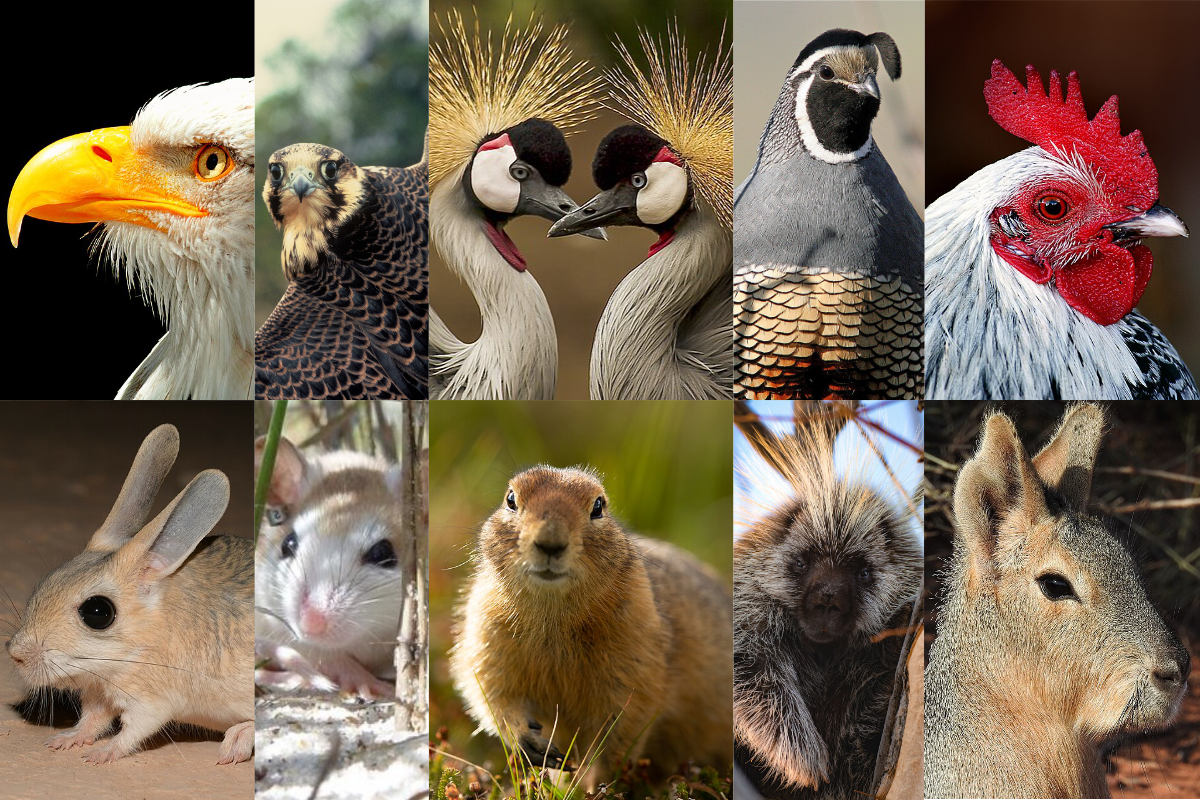
Faces are a vital part of every animal that hold both social and utilitarian significance. They are important for recognition of friend or foe, and many species have even developed evolutionary changes to their face based on their diet and eating habits. Beaks and snouts come in a vast array of forms, but, complex as faces are, the mechanism for developing facial morphology—the structure of the face—is even more intricate.
New research from Environmental Genomics and Systems Biology scientists Axel Visel and Laura Cook, in collaboration with the Max Planck Institute for Evolutionary Biology, describes how gene enhancers drive facial morphology. Enhancers are non-coding regions of genetic information that act as regulatory switches. Using chicken and mouse models, they mapped which genes are switched on—and which enhancers control those genes during facial development. These discoveries are catalogued in the VISTA Enhancer Browser, a freely available resource developed at Berkeley Lab that allows anyone to view enhancer data that has been validated in vivo.
While genes provide the basic building blocks for face development, it is changes in enhancers that likely drive differences in facial shape between birds and mammals. Enhancers increase modularity: the same gene can cause completely different outcomes based on which enhancers are present and active. “For example, if you want to make a beak slightly longer, say to reach nectar deep inside a flower, changing the underlying gene may accidentally break some other aspect of development, like the lungs,” explained Cook. “So, instead you can just change the instructions for when and where to turn a gene on or off.”
During embryonic development, specialized regions called developmental organizers send signals that direct cells to form specific structures. The frontonasal ectodermal zone (FEZ) is a developmental organizer responsible for determining the upper face in vertebrates. Conventionally, the FEZ has been defined by specific genes that are expressed in adjacent, mutually exclusive zones of the embryonic face. This work expands the definition by showing that, while there is exclusivity, the spatial domains are likely more flexible than previously thought, which may be a mechanism of facial diversity. It’s a new understanding that led to the discovery that instructions in mesenchymal cells—unspecialized connective tissue—show far greater variation between species than in the ectoderm.
The researchers also uncovered a novel signaling region in the nasal pit rim of mice. This nasal pit rim feature was absent in chickens and lizards, indicating that it could be a unique evolutionary development for mammals.
The research found that while genes are largely conserved between species, the regulatory sites are what vary most. “It’s not making new genes or even modifying the genes, it’s really focused on modifying the instructions,” said Cook. “That is the hypothesis: the way we generate facial diversity—or morphological diversity in general—is through changing these instructions that control where, when, and how much of a gene is expressed during development.” she added.
This means the discoveries from research into mice could be extrapolated to other mammals. For instance, in humans it may open the door to newly possible studies into the genetic basis of craniofacial differences such as cleft lip, cleft palate, and rare conditions, such as Treacher Collin’s syndrome.


