Nearly 100 years ago, the introduction of the phase-contrast microscope brought into clear view structures inside cells that had previously been too faint or washed out for biologists to study. The discovery revolutionized light microscopy and garnered a Nobel Prize in 1953.
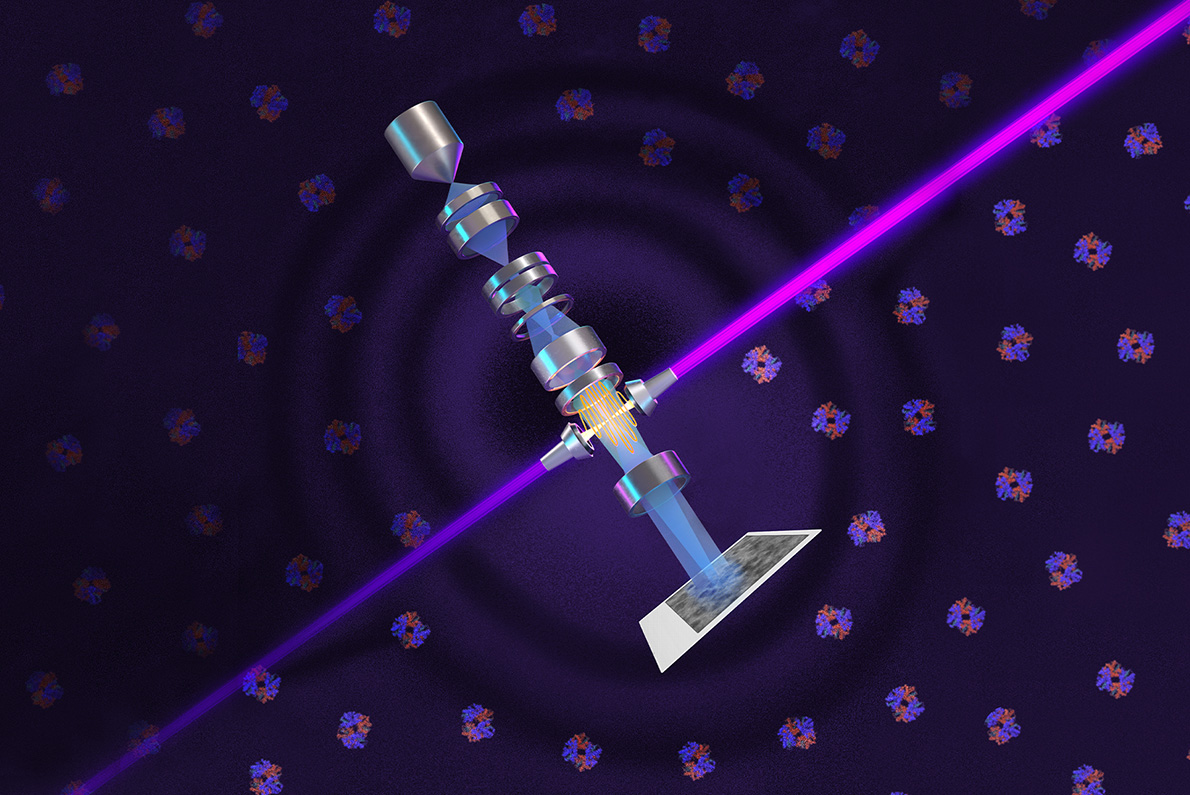
Now, a team of physicists including Molecular Biophysics and Integrated Bioimaging (MBIB) faculty scientist Holger Müller have adapted the phase-contrast technique to cryo-electron microscopy (cryo-EM), which has about 10,000 times the magnification of light microscopy. The technology employs a spot of laser light millions of times brighter than the Sun to convert invisible differences in the position of electron wave peaks and troughs—its “phase”—into visible differences in image contrast.
The new technology was brought to fruition by more than 15 years of theoretical and experimental work by leading microscopy scientists, collaboration with expert machinists, and support from Biohub. The phase plate is paired with a new, custom Thermo Fisher Scientific microscope that was developed to maximize the benefit of the plate’s ultra-bright laser. As reported in Science, resolution improved by up to 44% with the laser on, making details of small molecules that today’s best cryo-EM systems struggle to resolve clearer and sharper.
In their paper, Müller and his colleagues demonstrate the system’s power by imaging aldolase, a protein in muscle that is relatively easy to capture with today’s cryo-EM machines, and hemoglobin—a protein that carries oxygen in blood. Hemoglobin is a smaller protein that sits at the lower size limit for current machines and is often used as a benchmark for cryo-EM performance. The laser-phase plate improved the resolution of the protein structure in both cases, but more so for the hemoglobin.
Müller named the complete system “Theia” after the ancient Greek Titaness of light and radiance. “Before, studying structures with cryo-EM was like trying to look at paintings in a dark gallery. With Theia, it’s like the lights have been turned on for the first time,” he said.
Read more in the Berkeley Lab News Center.

