In the wake of radiation emergencies like nuclear accidents or radiological attacks, knowing whether individuals or populations were exposed to ionizing radiation is vital for managing medical treatment, protecting emergency workers and the public, and understanding the likelihood that any disease manifestations were induced by the incident. But existing technologies for screening large numbers of people for radiation require invasive blood collection, involve long processing times, and are ineffective at catching low doses of radiation exposure, which may still increase risk for developing cancer and other harmful health effects.
To address this gap, a team led by Antoine Snijders, a senior scientist and Head of the Bioengineering & Biomedical Sciences Department in the Biological Systems and Engineering (BSE) Division, developed a machine learning–powered pipeline that leverages the high sensitivity of infrared imaging as a part of Intelligence Advanced Research Projects Activity (IARPA) ’s Targeted Evaluation of Ionizing Radiation EXposure (TEI-REX) program. Changes in the composition and molecular structure of many biomolecules—such as DNA, lipids, proteins, and carbohydrates—are reliable reporters for exposure to ionizing radiation. When exposed to infrared radiation, the microscopic vibrations of these biomolecules produce spectral features that can reveal subtle shifts at the level of individual chemical bonds or functional groups. In particular, Fourier transform infrared (FTIR) spectroscopy, an imaging technique that measures how much light a given sample absorbs at different wavelengths across the infrared spectrum, is one of the most sensitive analytical tools currently available for this purpose.
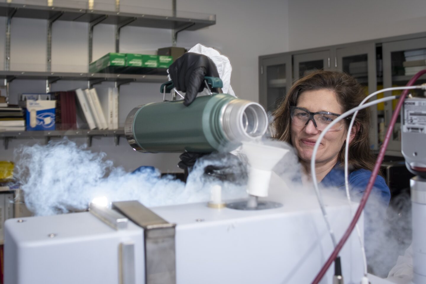
To use FTIR’s discriminatory power for studying radiation exposure, the researchers calibrated a microscopy setup for imaging and recording FTIR spectra of the skin on the outer ears of living mice. They then split the mice into two groups—one that was exposed to ionizing radiation, and one that was not—and repeatedly imaged the mice at different time points post-exposure. Drawing on AI as a second potent tool, the researchers tested whether various statistical machine learning models could pinpoint the spectral signatures of radiation exposure and predict whether or not an animal had been irradiated. They identified several models that were extremely competent at distinguishing which animals had been irradiated, even at super low doses for as long as three months after exposure.
“Detecting low-dose exposures 90 days post-irradiation is definitely pushing the envelope on what was believed to be possible,” said Jamie Inman, a BSE research scientist and first author on the study. This is in part because the skin cells of the mouse ear turn over roughly every eight to ten days. That the models could detect damage signatures so long after irradiation demonstrates that spectral imaging can capture biological changes in tissue imparted by low-dose radiation that persist long after the skin cells have renewed.
“Detecting low-dose exposures 90 days post-irradiation is definitely pushing the envelope on what was believed to be possible,” said Jamie Inman.
While this finding highlights the rich resolution of FTIR imaging and could reveal a more detailed picture of the stem cell and tissue microenvironment, it also points towards the viability of non-invasive, reagent-free, and real-time diagnostics for a host of biomedical conditions and applications. Noting that the machine learning models were effective even when trained on a surprisingly small sample size, the authors foresee that the approach could be adapted for use in human populations. They envision future deployable imaging devices that could probe the effects of ionizing radiation at a population scale. “This technology could improve awareness of intentional or accidental exposure events, improve protection of government personnel and uniformed service members, and support counter-proliferation efforts to improve national and global security,” Inman said.
Other Biosciences Area contributors to this work include: Environmental Genomics and Systems Biology (EGSB) graduate research assistant Yulun Wu and staff scientist Ben Brown; Molecular Biophysics and Integrated Bioimaging (MBIB) research scientist Liang Chen, staff scientists Corie Ralston and Peter Zwart, and senior scientist Hoi-Ying Holman; BSE research associate Ella Brydon, laboratory manager Kenneth Wan, senior scientist Jian-Hua Mao, and staff scientist Hang Chang.
Biosciences researchers also worked with collaborators in Berkeley Lab’s Accelerator Technology and Applied Physics Division (ATAP) including Kei Nakamura, Lieselotte Obst-Huebl, and Jared DeChant.


