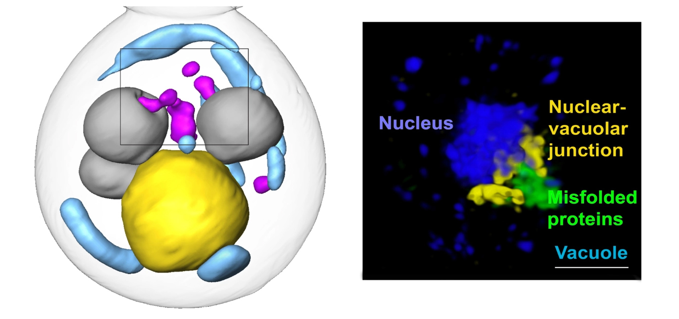
Left: A soft x-ray tomographic reconstruction of a yeast cell in which misfolded proteins (magenta) in the cytoplasm are being engulfed by vacuoles (gray), the cell’s protein degradation machinery. The cell nucleus is shown in yellow and mitochondria are shown in blue. Right: A super-resolution, visible-light microscopy reconstruction showing misfolded proteins (green) being extruded from the nucleus (blue) toward a vacuole through a nuclear-vacuolar junction (yellow). (Image credit: Fabián Morales-Polanco/Stanford University)

