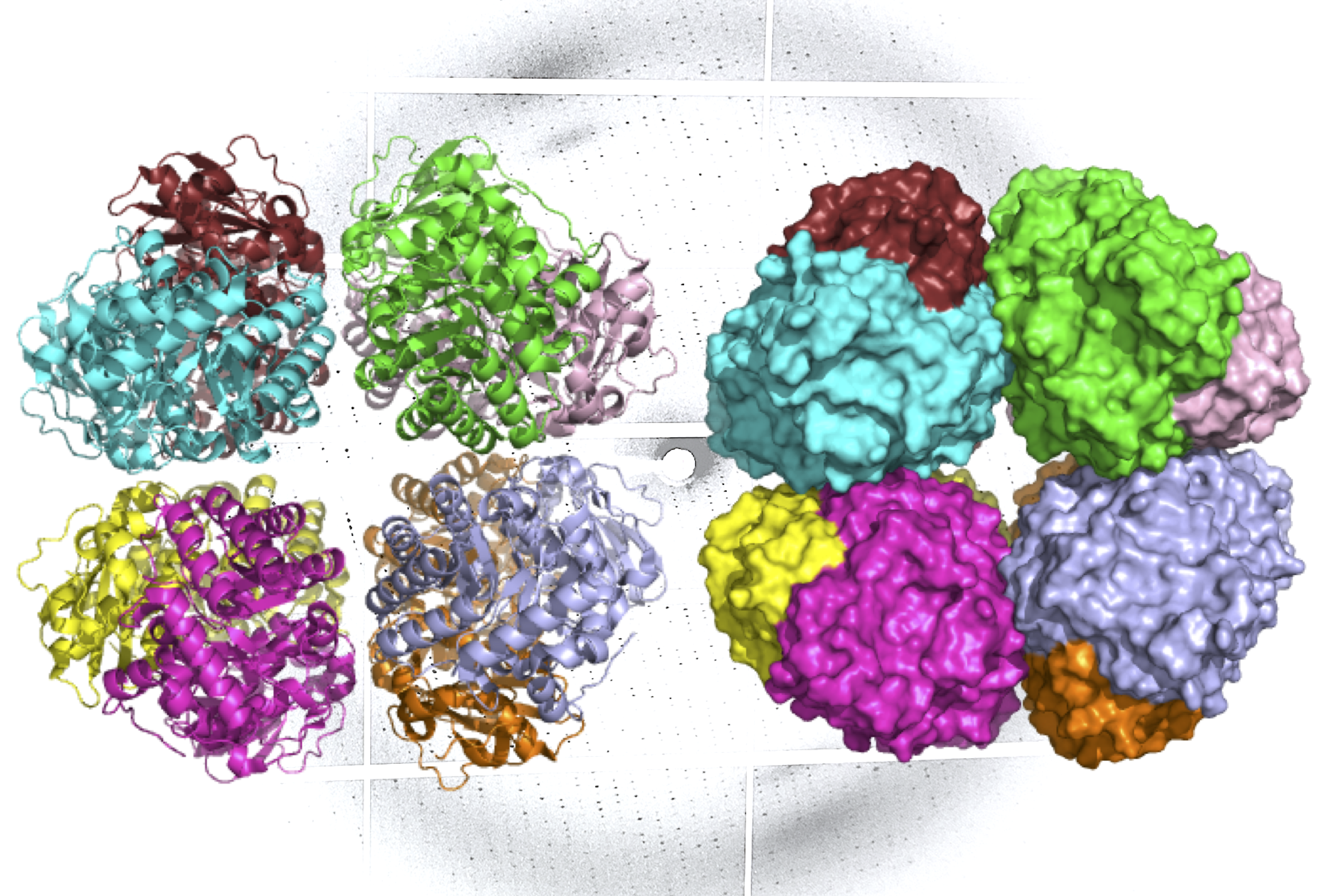
A ribbon diagram (L) and molecular surface representation (R) of carbon-fixing form I’ rubisco, showing eight molecular subunits without the small subunits. An x-ray diffraction pattern of the enzyme, also generated by the research team, is in the background. (Credit: Henrique Pereira/Berkeley Lab)

