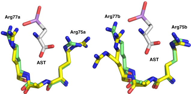
A conformational change of ArsN1 resulting from ligand binding. A portion of the AST binding site in ArsN1-AST (green) is superimposed with that of the unbound structure (yellow). Left and right cartoons depict chain A and chain B, respectively. Arg75a in chain A and Arg77b in chain B are closer to AST when substrate is bound.

