
Generate a mechanistic and predictive understanding of biological processes
Molecular Biophysics and Integrated Bioimaging (MBIB) Division investigators develop and apply molecular and meso-scale visualization and advanced spectroscopies to study biological processes, with the ultimate goal of enabling control, manipulation, and generation of biological function.
Much of the research that happens in the Division takes place within programs supported by various funders.
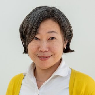
A senior scientist, Junko Yano is a co-principal investigator (co-PI) of the multi-institutional Liquid Sunlight Alliance (LiSA), a DOE Fuels from Sunlight Energy Innovation Hub. Yano is also a co-PI since 2022 of the Center for Electrochemical Dynamics and Reactions on Surface (CEDARS), a historically Black university–led DOE Energy Frontier Research Center.
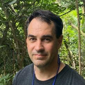
Greg Hura is a staff scientist working on the Structurally Integrated Biology for Life Sciences (SIBYLS) Beamline at the Advanced Light Source (ALS). He is also the Berkeley Lab lead for the Integrated Diffraction Analysis Technologies program and the principal investigator of Biopreparednesss Research Virtual Enviornment (BRaVE) Taskforce5.
Humphreys has worked in the University of California (UC) System for over 20 years gathering a wealth of experience and honing her analytical, communication, planning, and project management skills. She has a strong track record in management, having twice received the Berkeley Staff Assembly Excellence in Management Award.
Suzanne Baker is a senior scientist who studies aging and dementia using neuroimaging. Much of her focus has been on how to most accurately measure amyloid plaques and tau tangles, two proteins that are associated with Alzheimer’s disease. She currently is co-PI of a multi-center project on harmonization of tau positron emission tomography tracers. Her interests also include the effect of myelination, glymphatic clearance, neuroinflammation, and hormones on brain health.

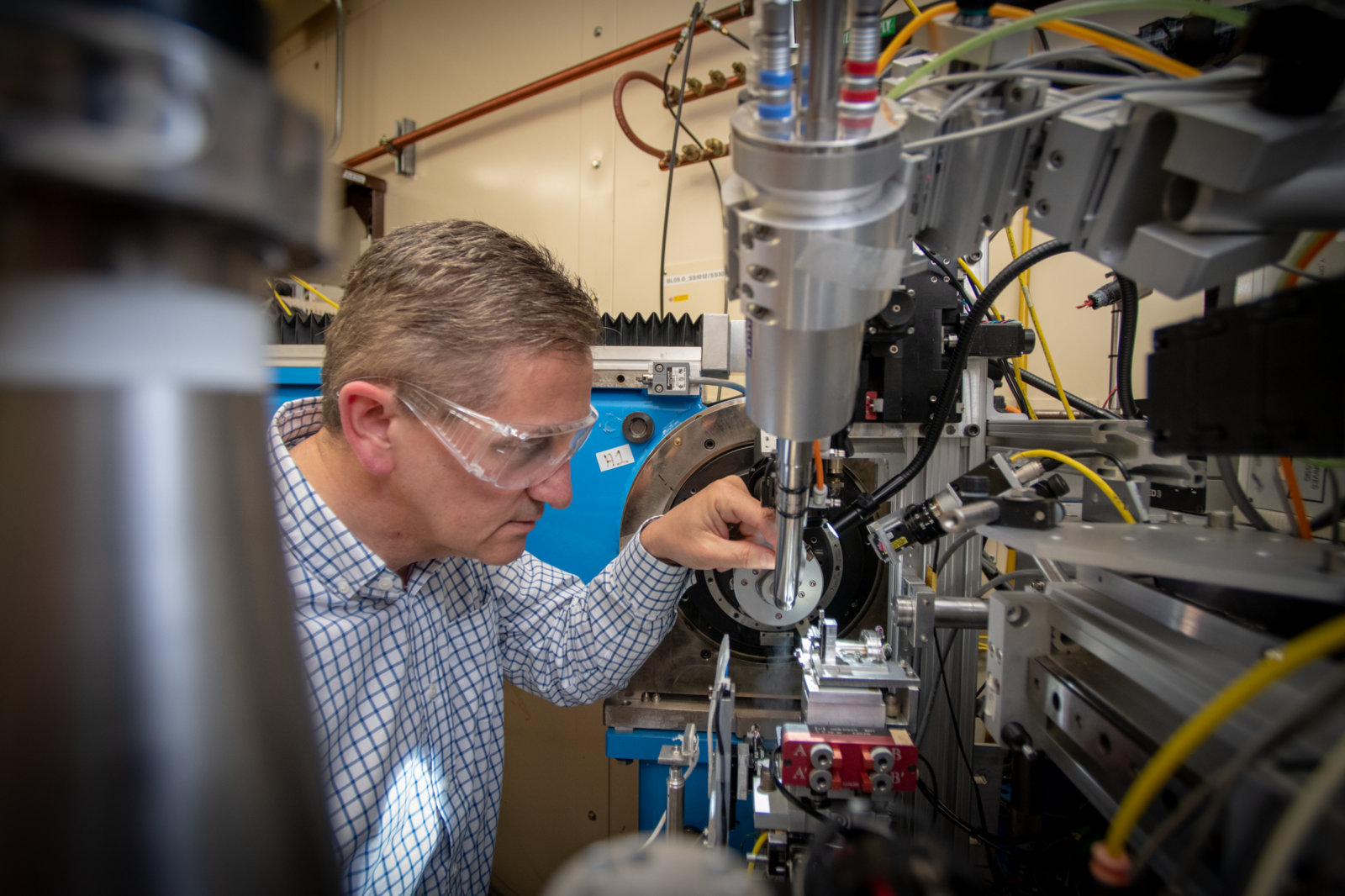
Jan Kern is a senior scientist and principal investigator of DOE and NIH funded projects focused on methods development for time-resolved X-ray studies of enzymes and on understanding the details of charge separation in natural photosynthesis. He regularly utilizes synchrotron and X-ray free electron laser facilities at Berkeley Lab and SLAC National Laboratory.

Specializing in small angle X-ray scattering (SAXS) and macromolecular crystallography, Susan Tsutakawa is a staff scientist at the SIBYLS beamline at the ALS. The goal of her research is to understand the structural mechanisms underlying protein function, with focus on proteins relevant to DNA and RNA processes in human cancer and biopreparedness. She works with CASP committee for experimentally testing protein structure predictions.
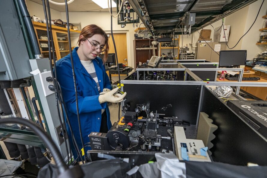
Bioenergetics Department investigators develop and apply imaging techniques to visualize atomic, molecular, and electronic structural level phenomena of biological systems—especially photosynthetic and bio-inspired synthetic systems that play a role in energy transfer and conversion.
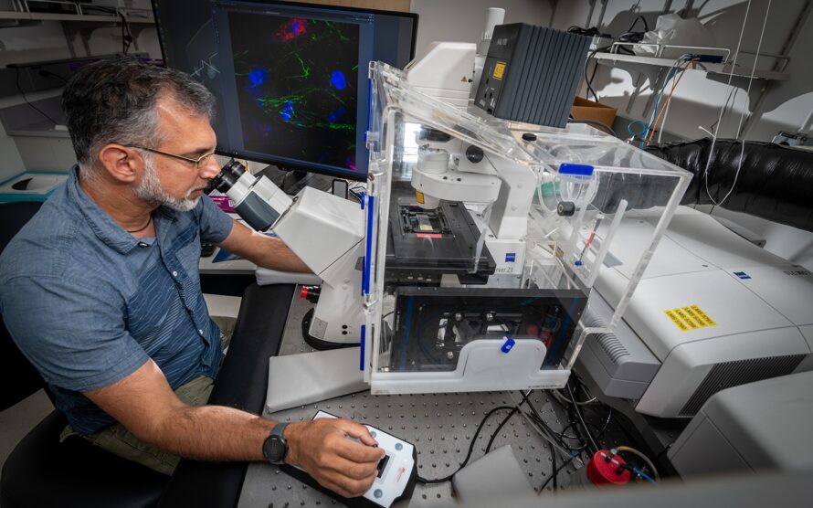
Cellular and Tissue Imaging Department investigators employ bioimaging at multiple scales and levels of spatiotemporal resolution, and integrate information obtained by the different imaging modalities to derive a comprehensive mechanistic understanding of physiology and diseases mechanisms.
Structural Biology Department researchers use a variety of techniques—including X-ray crystallography, X-ray free-electron laser crystallography and spectroscopy, electron cryo-microscopy, and X-ray tomography—to determine the molecular structures and gain dynamic information about macromolecules and investigate how changes in structure can affect their function.
We are committed to ensuring an open and welcoming workplace for all employees, contractors, affiliates, and visitors. This expectation applies to all roles and levels–from managers to supervisors to individual contributors–in the Division.

Scientists in the Biosciences Area are using X-ray technology at the Advanced Light Source to gain a better understanding of the structure of the COVID-19 virus’s molecular machinery. By mapping the structure of the proteins that the virus uses to infect cells, the researchers are providing critical information for other scientists working on drug and vaccine development.

