Bruce Cohen
Materials Staff Scientist

Divisions
Molecular Foundry
Secondary Affiliation:
Molecular Biophysics and Integrated Bioimaging
- Cellular and Tissue Imaging
Biography
Bruce Cohen, Ph.D. is a Staff Scientist at the Molecular Foundry and the Division of Molecular Biophysics & Integrated Bioimaging at Lawrence Berkeley National Laboratory in Berkeley, California (USA), where his research focuses on the design and synthesis of novel luminescent nanomaterials for bioimaging. He graduated with honors in Chemistry from Princeton and earned his Ph.D. in Chemistry and Biophysics from UC Berkeley working with Daniel E. Koshland, Jr. He earned a certificate in Neurobiology from Woods Hole Institute before working as a Howard Hughes Medical Association postdoctoral fellow in the laboratory of Lily Y. Jan (UCSF) developing organic and protein-based fluorescent probes for addressing problems in protein biophysics and bioimaging. He has a broad background covering nanoscience, biophysics, neuroscience, photophysics, and chemical biology. His current research interests include development of novel organic fluorophores for imaging and sensing, nanoparticle-based biosensors, and lanthanide-based upconverting nanoparticles as single molecule, quantitative, and deep tissue imaging probes.
Research Interests
Optical microscopy is the primary means of studying live cells in real time, enabling analysis of individual cellular components at high spatial and temporal resolution. Inorganic nanocrystals have shown promise as transformative probes for these imaging studies, with exceptional optical properties not found in other materials. Along these lines, we are developing novel nanocrystals as biosensors and single-molecule probes, improving bioconjugation and targeting chemistries, and imaging live cells with these reagents. We aim to integrate the development of novel luminescent nanomaterials into multidisciplinary efforts to address significant biological questions of cell function.
Imaging single molecules
Lanthanide-based upconverting nanoparticles (UCNPs) sum the energies of 2 incident NIR photons to emit one at visible wavelengths, an unusual property unlike anything found in the cell. UCNPs have significant advantages over other luminescent reporters, including an absence of on–off blinking, single-molecule multiphoton NIR excitation at powers approaching those used for standard one-photon confocal imaging, no overlap with cellular autofluorescence, and no measurable photobleaching under prolonged single-particle excitation. Our synthetic efforts have established control over UCNP size to produce smaller nanocrystals more compatible with many imaging applications. Current efforts are aimed at optimizing single nanocrystal brightness for extended single-molecule imaging in live cells, and developing UCNPs capable of chemical sensing for studying cellular biochemistry.
Optical biosensors
Many nanocrystals exhibit brightness and stability far superior to conventional fluorophores, making them ideal as the bases for biosensing reagents. We have developed quantum dot and UCNP energy transfer-based systems for the sensitive detection of cellular chemistry and protein motions. Our current work focuses on developing bright, selective sensors for studying neuronal activity, including voltage sensors, sensors of neurotransmission in the brain, and ion sensors.
Nanocrystal biocompatibility and targeting
Broadening the scope of nanocrystal surface conjugation chemistry is essential to expand their reach for imaging applications. High-quality UCNPs and quantum dots are synthesized in hydrophobic solvent and must be transferred to water and made biocompatible to have any utility as imaging probes or biosensors. An ongoing challenge in applying nanocrystals as probes for cellular imaging is improving their aqueous passivation and developing new reactions that work on nanocrystal surfaces. A growing area of research for us is development of new bioconjugation chemistries, including click reactions, SpyCatcher ligation, and bioorthonganol reactions for immunotargeting.
Recent Publications
Related News
Congratulations to Biosciences Area Director’s Award Recipients
Each year, the Berkeley Lab Director’s Achievement Award program recognizes outstanding contributions by employees to all aspects of Lab activities. Several Biosciences Area personnel are among the 2025 honorees.
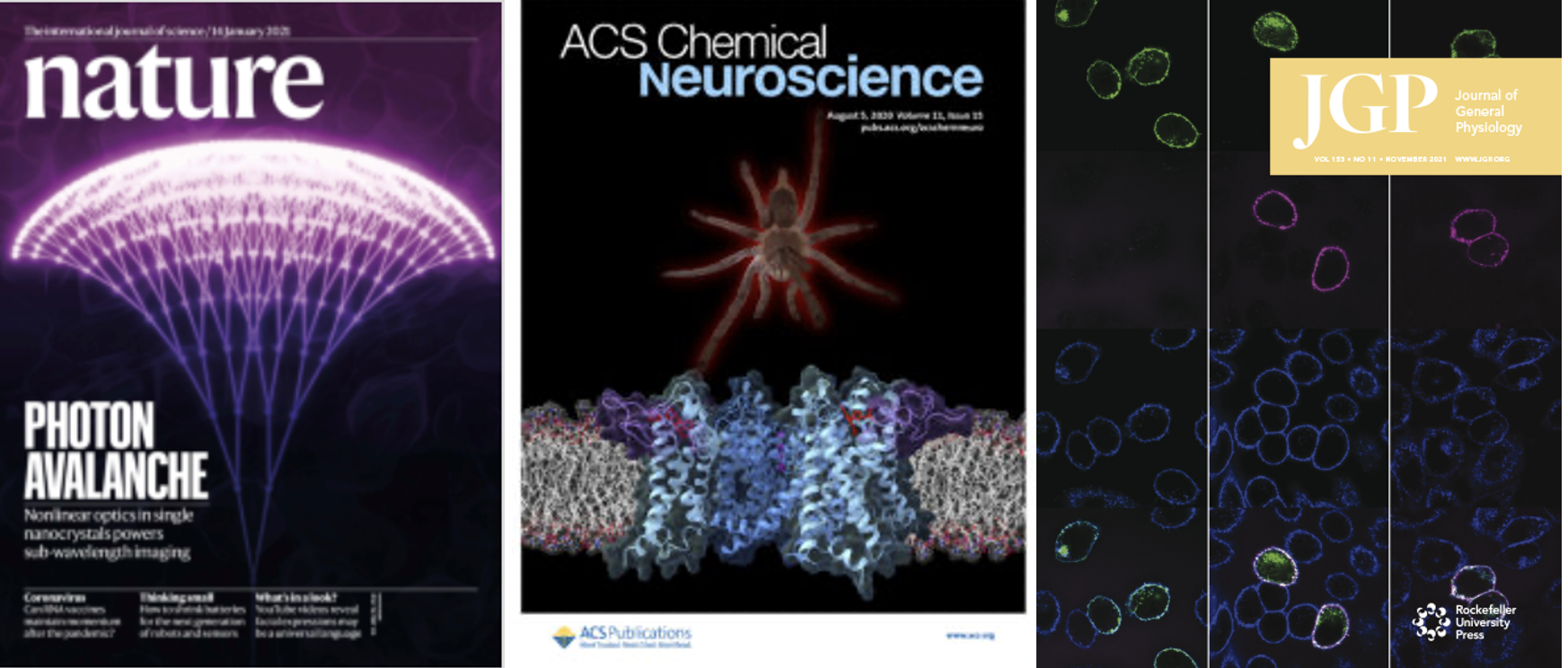
Shine On: Avalanching Nanoparticles Break Barriers to Imaging Cells in Real Time
The diffraction limit is a fundamental property of light that has long prevented optical microscopes from bringing into focus anything smaller than half the wavelength of visible light (~200 nanometers), which is at least an order of magnitude larger than the tiny protein machines that keep cells, and us, running. A team of researchers co-led scientists in Berkeley Lab's Molecular Foundry and Columbia University’s school of engineering developed a new class of crystalline material that, when used as a microscopic probe, overcomes the diffraction limit without heavy computation or a super-resolution microscope. The amazing new material, called avalanching nanoparticles (ANPs), will advance high-resolution, real-time bio-imaging of a cell’s organelles and proteins, as well as the development of ultrasensitive optical sensors and neuromorphic computing that mimics the neural structure of the human brain, among other applications. The work was reported in a cover article in the journal Nature.

