Advanced Microscopy Facility
- Introduction
- Getting Access
- Usage Charges & Supplies
- EHS Information
- Data Storage and Analysis
- Zeiss LSM710 Confocal Laser Scanning Microscopes
- DeltaVision OMX Super-Resolution Light Microscopy System
- Zeiss Multispectral Scanning Microscope
- Zeiss Axiovert119 Epifluorescence Microscope
- Zeiss Spinning Disk Confocal and Wide-field Epifluorescence Microscope
- Cellomics Arrayscan
- Resources
- Contact Us
Introduction
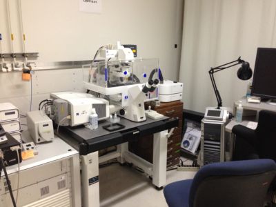
The mission of the Advanced Microscopy Facility is to support biosciences research at Berkeley Lab by providing capabilities to which individual laboratories might not otherwise have access. Others interested in using the facility please see below under “Getting Access.” Instruments in the facility include two Zeiss LSM710 confocal laser scanning microscopes, a DeltaVision OMX super-resolution light microscopy system, a Zeiss multispectral scanning microscope, a Zeiss Axiovert119 epifluorescence microscope, a Zeiss spinning disk confocal and wide-field epifluorescence microscope, and a Cellomics Arrayscan high-content imaging system.
The facility is managed by David Acevedo. For access, training, and questions, please contact him; he can be reached via phone at (510) 486-6139 and via e-mail.
Getting Access
The Advanced Microscopy Facility is available to all Berkeley Lab researchers with a Berkeley Lab employee or affiliate appointment; a valid Berkeley Lab project ID is required for the usage charges. Others interested in using the facility, please request access via the Science Exchange website or contact David Acevedo.
The facility is located at 717 Potter Street, Berkeley. Training is required before use of the instruments. Training sessions can be scheduled by emailing David Acevedo. Relevant Environment/Health/Safety (EHS) training also needs to be completed prior to access.
Once trained, users can sign up for and use the instruments for which they completed training. Sign up through the Facility Online Manager (FOM) system, selecting “Lawrence Berkeley Laboratory Cytometry/Microscopy” facility. For instructions on the FOM system see this document; and refer to this document on how to add this scheduling tool to a Google or Outlook calendar.
In order to log in on the computers that control each instrument, an account is needed on Berkeley Lab’s Active Directory or Windows Domain system (Note: This is different from the LDAP account which is used for e-mail, LETS, etc.). Many Berkeley Lab users already have an account and can simply use that to log in. If not, visit this website to request a Windows Domain account. More information about Active Directory.
Read the section below for information about usage charges and supplies.
Usage Charges & Supplies
The charge for the use of any instrument is $29.00 per hour. Also, if operator assistance is needed an additional fee of $120.00 per hour is charged (Fee schedule). For external users, additional charges will apply; please use the Science Exchange website for access to our facility and to request a quote.
Users are charged via the Facility Online Manager (FOM) system. Please download and read the instructions for setting up an account.
The Advanced Microscopy Facility provides immersion oil, double distilled water, lens cleaning solution and lens paper. All other supplies must be brought by the users, which includes slides, transfer pipettes, gloves. In the resources section below is a list of the typical supplies and the ordering information.
EHS Information
The Advanced Microscopy Facility is located in a BSL-2 lab; no food or drinks are allowed. Each user must have completed and must be current on the following Environment/Health/Safety (EHS) training:
- EHS0735/738 Bloodborne Pathogen training and annual refresher;
- EHS0745 Hepatitis B Medical Surveillance training;
- EHS0604 Hazardous Waste Generator training;
- EHS0730 Biohazardous Waste training.
Visit the Berkeley Lab training website for more information.
The use of the facility and the method of transport of samples between the home lab and the facility must be described in the Work Planning and Control documentation of the home lab’s Principal Investigator. If you have any questions, please contact David Acevedo for information.
Data Storage and Analysis
The computers that control each instrument may temporarily be used to store acquisition data; these data will be kept for one week after acquisition. Users are responsible for their own data storage and can copy it over the network to their lab’s server, copy it to a USB memory stick or portable USB hard drive, or burn it onto a CD or DVD.
Zeiss LSM710 Confocal Laser Scanning Microscopes
The Zeiss LSM710 is a top of the line laser scanning confocal microscope incorporating a Zeiss Observer Z1 inverted microscope body. The microscope uses a fully automated stage, objective selector, filter cube and autofocus. The system is outfitted with four lasers supplying six discreet laser lines for excitation (405nm, 458nm, 488nm, 514nm, 561nm, 633nm) and uses three photomultiplier tubes as detecters, incorporating one 32-element spectral detector. The optical collection uses advanced spectral scattering elements to define wavelength selection for optimized multicolor imaging. The Zeiss system is computer-controlled using Zeiss ZEN software, which handles all aspects of image excitation and acquisition, and is capable of single track multicolor and multi-track single/multi-color imaging. The confocal pinhole is computer-controlled helping to optimize pinhole to NA of objective and emission wavelength producing crisp section images. With installed high NA water and oil immersion objective lenses the system is capable of producing high definition 3-dimensional images of cellular organelles and structures. Available modes include Z-stack, FRAP, Time course, Tiles, Regions, Color Deconvolution. LSM710#1 also incorporates an integrated live cell chamber providing a 37C, 5% CO2 humidified environment for extended live cell imaging.
LSM710#2 is exactly the same but does not include a live cell chamber.
DeltaVision OMX Super-Resolution Light Microscopy System
The DeltaVision OMX super-resolution light microscopy system surpasses previous resolution limits and allows imaging beyond the surface of the coverslip with multiple probes. The system’s structured illumination technology images more of the biology and resolves features invisible with traditional microscopy. Using the DeltaVision OMX, you can image from five to twenty microns into cells and tissues.
The GE OMX super-resolution light microscope is a state of the art instrument capable of resolving fluorescent structures to within 100 nanometers of each other. The system is capable of Structured Illumination (SIM), Total Internal Reflection Fluorescence (TIRF) and PALM-STORM fluorescence imaging. With these techniques resolutions of 100-150 nanometers can be achieved using fluorescent probes. The system is outfitted with four lasers supplying four discreet laser lines for excitation (405nm, 488nm, 561nm, 635nm). It is equipped with a computer controlled stage for precise positioning. The system comes with three very high NA objective lenses for high resolution imaging. The system is computer controlled using API software derived from Delta Vision microscope systems. An integrated workstation provides software algorithms for image deconvolution and reconstruction.
Zeiss Multispectral Scanning Microscope
The Zeiss multispectral scanning microscope incorporates a Zeiss AxioSkop2 upright microscope body matched to a CRI multispectral scanning system. The system is a combination of hardware and software. The patented liquid crystal (LC) tuning element functions as a high-quality interference filter that enables the transmitted light to be electronically tunable. This allows rapid, vibrationless selection of wavelengths in the visible or NIR range, and digital images (called image cubes) are captured at the specified wavelengths. The intuitive Nuance acquisition and analysis software performs spectral classification and unmixing of overlapping dyes or stains that may look indistinguishable to the naked eye but have differing spectral signatures. Image cubes consist of standard 8-bit or 12-bit Tagged-Image File Format (TIFF) files in order to maintain compatibility with popular third-party image processing programs. Other file formats are also supported for saving individual images in color or grayscale and for exporting to other more specialized image processing programs.
Zeiss Axiovert119 Epifluorescence Microscope
The Zeiss Axiovert119 is a conventional epifluorescence microscope based on the Zeiss Axiovert 135TV inverted microscope body. It has a semi-automated stage and excitation optics. It is equipped with a X-Cite closed loop fluorescent lamp source and multiple sets of conventional multipass emission filters for multicolor imaging. The microscope is equipped with a single sensitive Axiocam digital camera connected to a computer running Zeiss Axiovision software for image acquisition.
Zeiss Spinning Disk Confocal and Wide-field Epifluorescence Microscope
The Zeiss spinning disk confocal and wide-field epifluorescence microscope is a duel function microscope based on a Zeiss Axiovert 200M inverted microscope body. It can be configured either with a Hamamatsu CCD camera or Solamere Spinning Disk option. In the wide-field mode fluorescence excitation is provided by a X-Cite closed loop lamp system. It is configured with a standard set of filter cubes to optimally observe blue fluorescence (DAPI), green fluorescence (FITC), red fluorescence (Texas Red) and near infrared (Cy5) fluorescence.
When the spinning disk confocal option is attached, excitation is provided by a 405nm laser, a 488nm laser, a 561nm laser and a 645nm laser. The spinning disk body is mated to a Stanford Photonics XR/Mega-10 ECCD camera for low light level imaging. The stage is contained in a closed environment chamber maintaining constant 37C temperature and 5% CO2 humidified air for long time lapse imaging of live cells. Spinning disk confocal technology can provide high resolution 3D imaging over long time courses with little to no photon damage to cells due to the spinning disk configuration.
Cellomics Arrayscan
The Thermo Scientific Cellomics® ArrayScan® VTI HCS Reader is a modular, high-content screening instrument designed for high-capacity automated fluorescence imaging and quantitative analysis of fixed and live cells. The instrument features optics by Carl Zeiss®, a broad white-light source, a scientific grade digital camera, and integrated acquisition and analysis software.
Fully automated and software-controlled compatible, with samples in SBS-compliant microwell plates and microscope slides. Hamamatsu ORCA®-ER cooled 12 bit grayscale digital CCD camera offering high-resolution, 1344 x 1024 pixel array with 6.45µm x 6.45µm pixel size. Automated objective changer giving increased resolution and sensitivity.
Resources
Fee Schedule
Facility Online Manager (FOM) system account set-up instructions
Facility Online Manager (FOM) system – how to add to Google or Outlook calendar?
Google Drive Microscopy Folder, containing instrument manuals, user guides, etc.
Contact Us
If you have any questions about the Advanced Microscopy Facility, please contact David Acevedo via phone at (510) 486-6139 or e-mail.