- Introduction
- Getting Access
- Usage Charges & Supplies
- EHS Information
- Data Storage and Analysis
- Zeiss LSM710 Confocal Laser Scanning Microscopes
- Resources
- Contact Us
Introduction
The mission of the Advanced Microscopy Facility is to support biosciences research at Berkeley Lab by providing capabilities to which individual laboratories might not otherwise have access. There is one Zeiss LSM710 confocal laser scanning microscope (LSM2), which is managed by Antoine Snijders. For access, training, and questions, please contact him via email or phone (see Contact Us, below).
Getting Access
The Advanced Microscopy Facility is available to all Berkeley Lab researchers with a Berkeley Lab employee or affiliate appointment; a valid Berkeley Lab project ID is required for the usage charges.
The facility is located at 717 Potter Street, Berkeley. Training is required before use of the instrument. Training sessions can be scheduled by emailing Antoine Snijders. Relevant Environment/Health/Safety (EHS) training also needs to be completed prior to access.
In order to log in on the computers that control each instrument, an account is needed on Berkeley Lab’s Active Directory or Windows Domain system (Note: This is different from the LDAP account which is used for email, LETS, etc.). Many Berkeley Lab users already have an account and can simply use that to log in. If not, visit this website to request a Windows Domain account. More information about Active Directory.
Read the section below for information about usage charges and supplies.
Usage Charges & Supplies
The charge for the use of any instrument is $29.00 per hour. Also, if operator assistance is needed an additional fee of $120.00 per hour is charged (Fee schedule).
The Advanced Microscopy Facility provides immersion oil, double distilled water, lens cleaning solution and lens paper. All other supplies must be brought by the users, which includes slides, transfer pipettes, gloves. In the resources section below is a list of the typical supplies and the ordering information.
EHS Information
The Advanced Microscopy Facility is located in a BSL-2 lab; no food or drinks are allowed. Each user must have completed and must be current on the following Environment/Health/Safety (EHS) training:
- EHS0735/738 Bloodborne Pathogen training and annual refresher;
- EHS0745 Hepatitis B Medical Surveillance training;
- EHS0604 Hazardous Waste Generator training;
- EHS0730 Biohazardous Waste training.
Visit the Berkeley Lab training website for more information.
The use of the facility and the method of transport of samples between the home lab and the facility must be described in the Work Planning and Control documentation of the home lab’s Principal Investigator. If you have any questions, please contact Antoine Snijders for information.
Data Storage and Analysis
The computers that control each instrument may temporarily be used to store acquisition data; these data will be kept for one week after acquisition. Users are responsible for their own data storage and can copy it over the network to their lab’s server, copy it to a USB memory stick or portable USB hard drive, or burn it onto a CD or DVD.
Zeiss LSM710 Confocal Laser Scanning Microscope
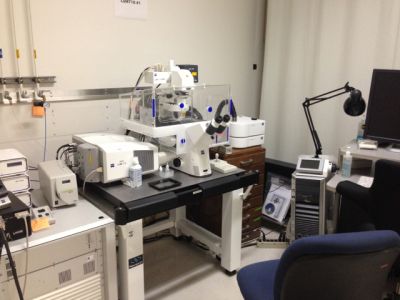
The Zeiss LSM710 is a top of the line laser scanning confocal microscope incorporating a Zeiss Observer Z1 inverted microscope body. The microscope uses a fully automated stage, objective selector, filter cube and autofocus. The system is outfitted with four lasers supplying six discreet laser lines for excitation (405nm, 458nm, 488nm, 514nm, 561nm, 633nm) and uses three photomultiplier tubes as detecters, incorporating one 32-element spectral detector. The optical collection uses advanced spectral scattering elements to define wavelength selection for optimized multicolor imaging. The Zeiss system is computer-controlled using Zeiss ZEN software, which handles all aspects of image excitation and acquisition, and is capable of single track multicolor and multi-track single/multi-color imaging. The confocal pinhole is computer-controlled helping to optimize pinhole to NA of objective and emission wavelength producing crisp section images. With installed high NA water and oil immersion objective lenses the system is capable of producing high definition 3-dimensional images of cellular organelles and structures. Available modes include Z-stack, FRAP, Time course, Tiles, Regions, Color Deconvolution.
Resources
Fee Schedule
Google Drive Microscopy Folder, containing instrument manuals, user guides, etc.
Contact Us
For questions pertaining to LSM2, please contact Antoine Snijders via email or phone at (510) 486-7235.

