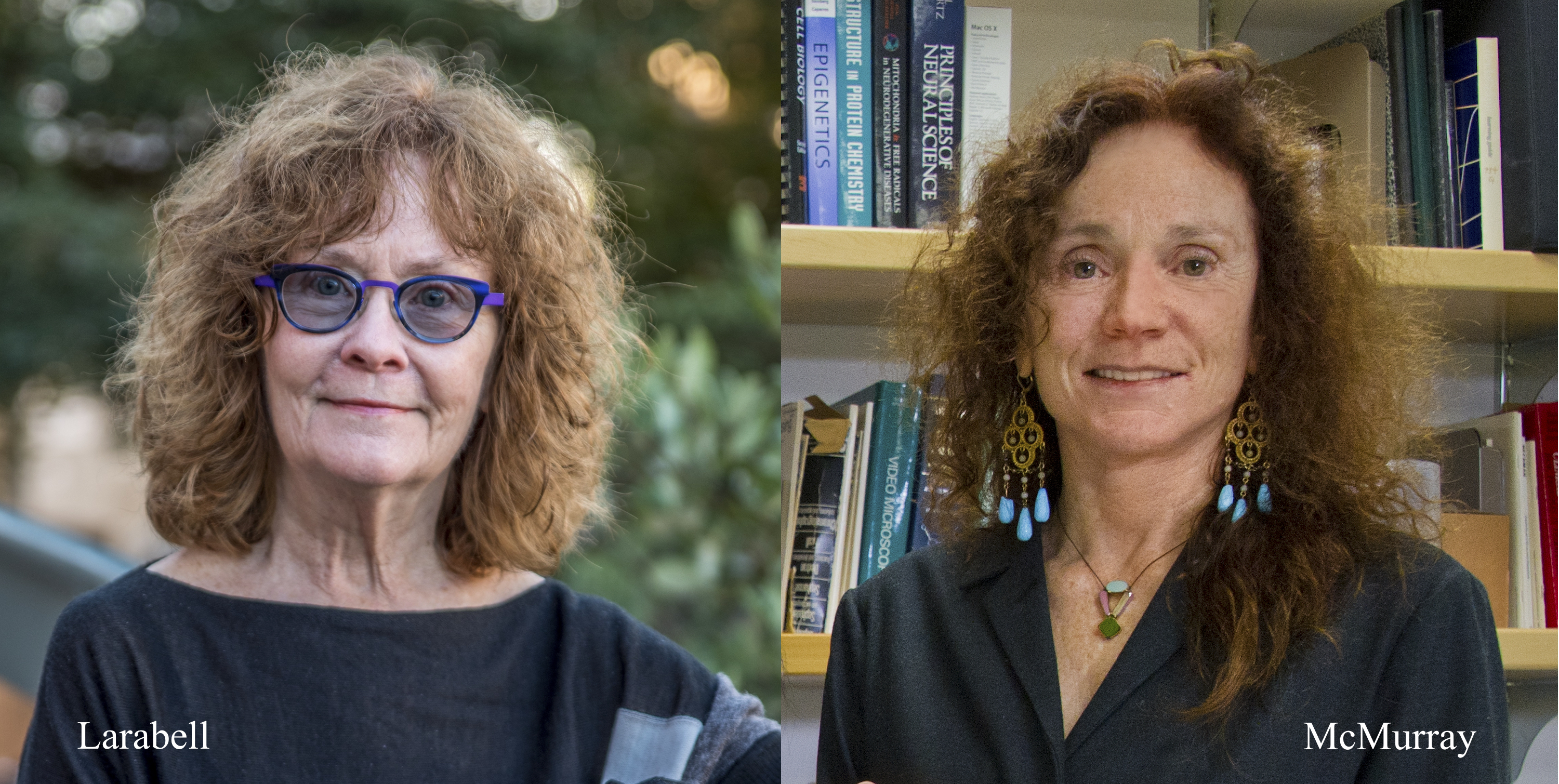
Two proposals, from Biosciences Area researchers Cynthia McMurray and Carolyn Larabell, have been recommended for funding by the Chan Zuckerberg Initiative (CZI) through a new grant program focused on developing collaborative computational tools to support the Human Cell Atlas (HCA). A global endeavor governed by an organizing committee co-chaired by Aviv Regev at the Broad Institute of MIT and Harvard and Sarah Teichmann at the Wellcome Trust Sanger Institute, the HCA will generate a variety of molecular and imaging data across a range of modalities and spatial scales. To advance the analysis, interpretation, and dissemination of these data, the CZI issued an open call in July 2017 for proposals aimed at creating new computational tools, algorithms, visualizations, and benchmark datasets. After a thorough evaluation process, CZI recommended 85 projects (out of nearly 300 submissions) for funding, and will provide $15 million in total over one year for their execution.
 McMurray, a senior scientist in the Molecular Biophysics and Integrated Bioimaging (MBIB) Division, plans to develop a non-destructive and label-free methodology, spectral phenotyping, to identify a cell type by its spectral properties. Using signals from IR, Raman, and fluorescence, a machine image-based algorithm will “learn” what features identify a cell type—a method conceptually akin to how “face recognition” software works. The overarching goal is to rapidly identify a cell type from complex mixtures or heterogeneous tissues, isolate it, and match it to a phenotype in control and disease states. The specificity and sensitivity make the technique an excellent tool for linking cell structure and function in vivo. This grant to McMurray was made possible through the Berkeley Lab Foundation.
 Larabell, a professor at the UC San Francisco School of Medicine and a faculty scientist in MBIB, plans to integrate machine-learning algorithms for segmentation into the pipeline for processing soft X-ray tomography (SXT) cell imaging data. SXT rapidly visualizes mesoscale functional structures (e.g. organelles, molecular machines) of intact, unperturbed cells better than any other imaging technique. However, segmenting and annotating the tomographic reconstruction remains a labor-intensive manual process. Using supervised machine learning to train computers to automatically identify and isolate each of the major organelles from other cell contents will overcome this bottleneck. In addition, giving the task of segmentation over to computers will ensure objectivity and consistency of data deposited in the Human Cell Atlas.
“We are excited to begin working on these promising projects with new partners from across the globe,” said Cori Bargmann, Head of Science for the CZI. “These grantees include experts in experimental biology, engineering, and computational biology. Enabling them to collaborate and bring their diverse perspectives to the work is the core of our approach to advancing biomedical science.”


