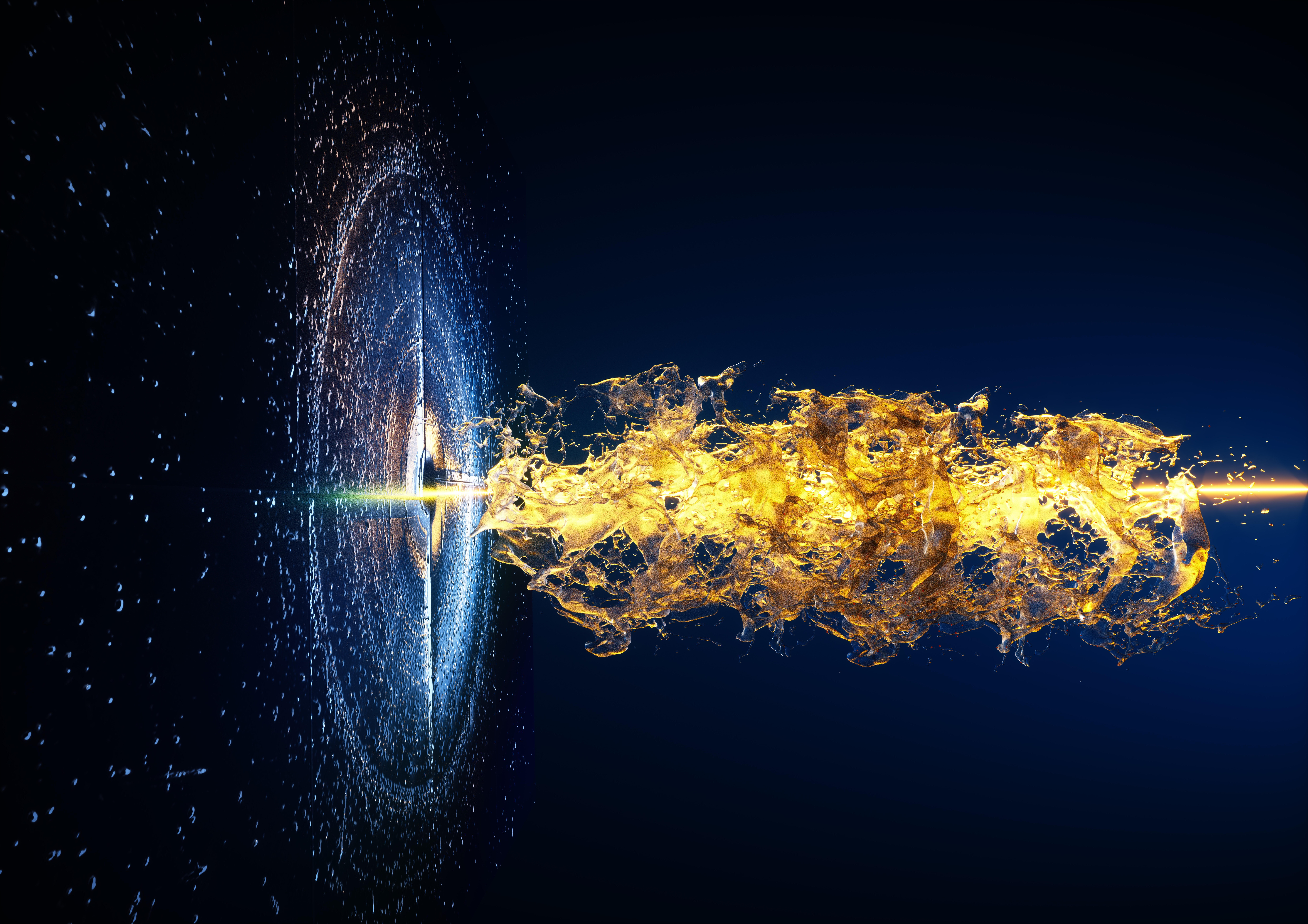
Studying materials at an atomic level allows us to understand and push the boundaries of what’s possible in science—from drug development to enabling breakthroughs in nanotechnology. Aaron Brewster, a staff scientist in the Molecular Biophysics and Integrated Bioimaging (MBIB) Division has long been interested in using X-ray crystallography to investigate the structure and inner workings of materials and proteins.
In 2022, Brewster was invited to Tokyo, Japan, as part of a larger collaborative effort to explore a new technique within X-ray crystallography that shrinks the time and cost it typically takes to investigate a material’s structure. Their scientific breakthroughs and the trials and tribulations of their work abroad are highlighted in a new short documentary, “BEAMTIME: Crystal Hitters.”
Brewster joined Berkeley Lab in 2013. In recent years his research has focused on developing computer algorithms that help decode the data generated in this field of study. In 2014, he helped develop an algorithm that deciphered crystallography data from Alzheimer disease–related research and, years later, catalyzed a separate conversation with a group of researchers at the University of Connecticut looking to do analogous experiments with small molecules.
“The structure lets us be guided to the function. And when we understand the function, we can understand how to change the structure, to give it a new function,” Brewster said.
Whereas usually X-ray crystallography requires that the material in question be coaxed into a large crystallized form (which not all substances tolerate), this new technique—known as small molecule serial crystallography—requires only a powder version of the material. “It turns out that the powder is easier to make and still provides information about the atomic structure,” Brestwer said. “It blows open the door on the number of different kinds of materials that we can study.”

For Brewster, being involved in this large-scale, collaborative experiment abroad was challenging and rewarding in equal measure. There were roughly a dozen people working together: to prepare the samples, work on the software, run the beamline, and solve the structures. “When things aren’t working according to plan, it’s stressful,” Brewster said. “But then when everything starts to click and it all falls into line, it’s glorious.”
J. Nathan “Nate” Hohman, assistant professor of chemistry at the University of Connecticut and lead scientist on this work, is now turning his focus to publishing the library of materials solved on this trip. And back at Berkeley Lab, Brewster is working to understand how this new technique might be adapted and made available at the Advanced Light Source synchrotron while colleagues are investigating how AI technology might further accelerate breakthroughs.


