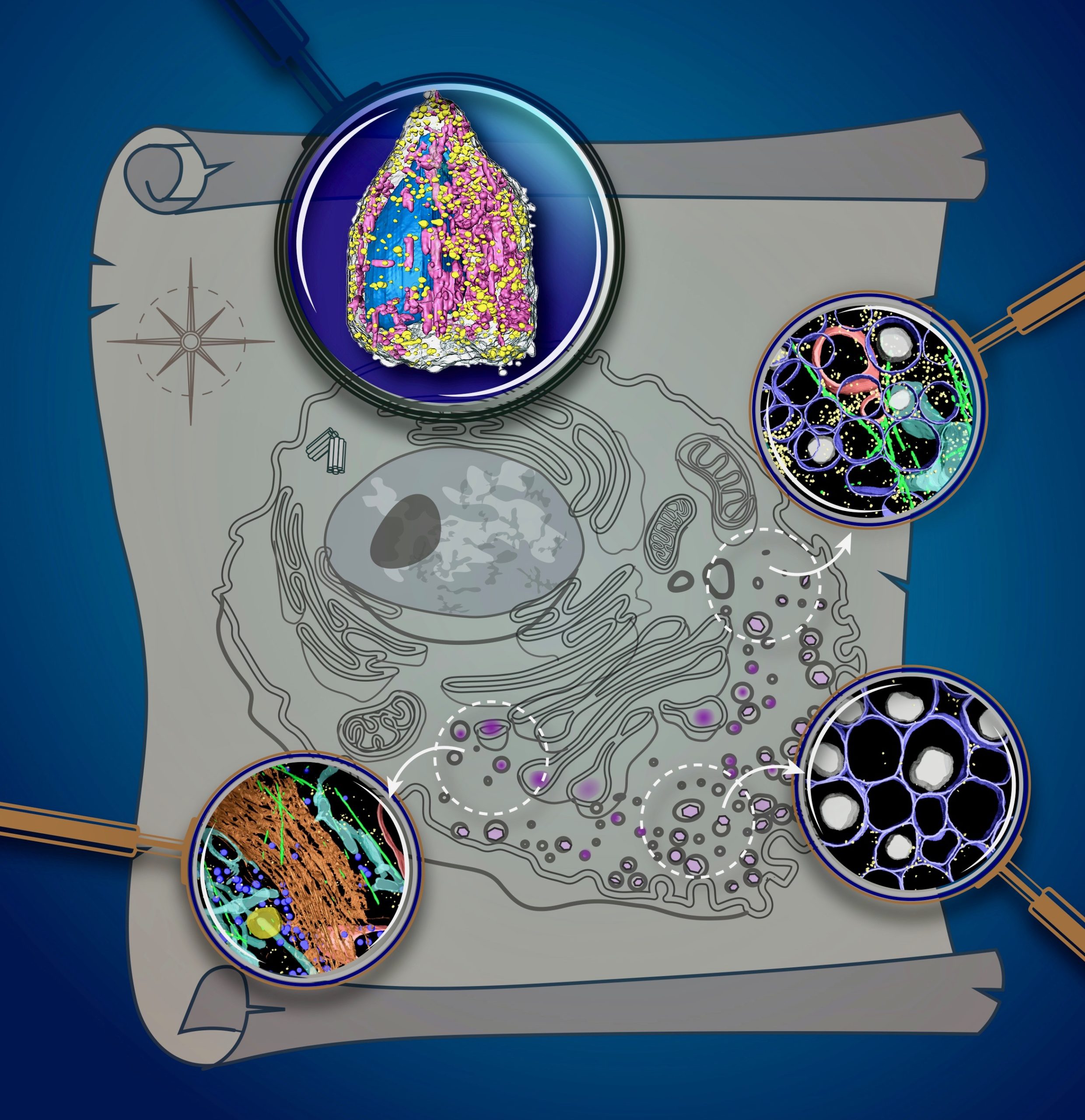
To fully understand cells, we need to characterize the structures that make them up, and to identify their contents. Thanks to advanced imaging technologies, scientists have examined many different components of cells; some current approaches can even map the structure of these molecules down to each atom. However, getting a glimpse of how all these parts move, change, and interact within a dynamic, living cell has always been a grander challenge.
A team based at Berkeley Lab’s Advanced Light Source is making waves with its new approach for whole-cell visualization, using the world’s first soft X-ray tomography (SXT) microscope built for biological and biomedical research. In its latest study, published in Science Advances, the team used its platform to reveal never-before-seen details about insulin secretion in pancreatic cells taken from rats. This work was done in collaboration with a consortium of researchers dedicated to whole-cell modeling, called the Pancreatic β-Cell Consortium.
Carolyn Larabell, Director of the National Center for X-ray Tomography (NCXT) and a Berkeley Lab faculty scientist in the Molecular Biophysics and Integrated Bioimaging (MBIB) Division, was the lead author on the study.
Read more in the Berkeley Lab News Center.


