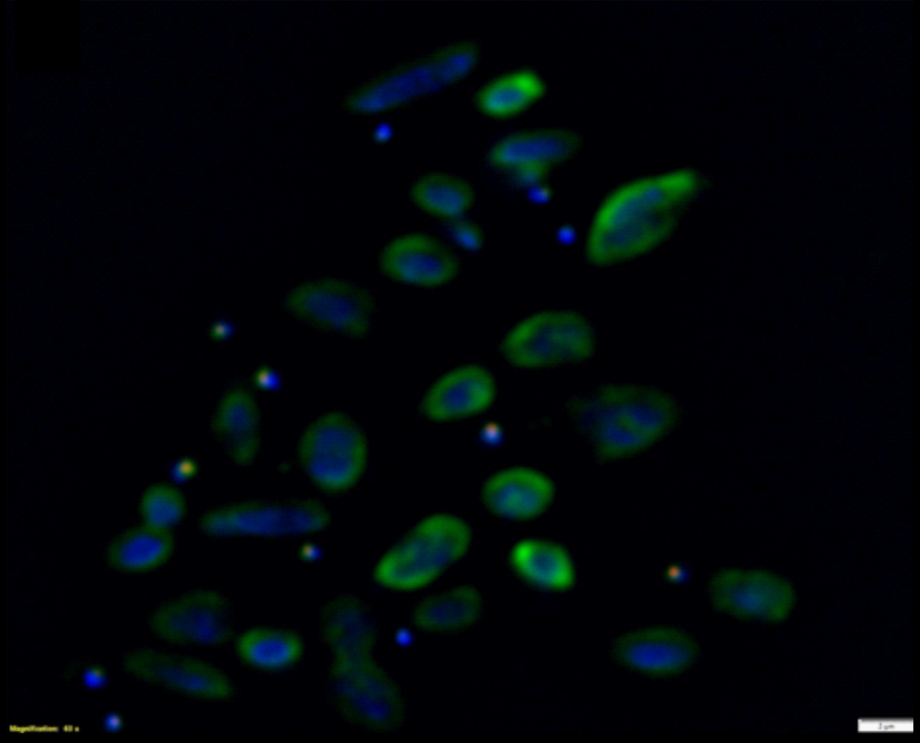
FISH of Nha-C enrichment with Hrr. lacusprofundi ACAM34-hmgA. Fluorescence micrograph shows individual Nha-C cells amongst Hrr. lacusprofundi cells. Nha-C cells labelled with a Cy5 (red fluorescence) conjugated probe; Hrr. lacusprofundi cells labelled with a Cy3 (yellow fluorescence, recolored to green to improve contrast) probe; all nucleic-acid containing cells stained with DAPI (blue fluorescence). Composite image of all three filters. Scale bars represent 2 µm. (Josh Hamm, UNSW)

