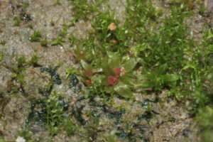
The structure of photosystem I is from the moss Physcomitrella patens. (Credit: HermannSchachner [CC0], Wikimedia Commons)
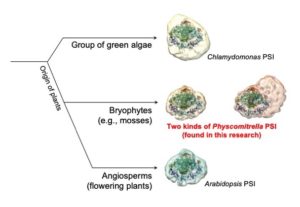
The team found that in this moss the structure of this complex of several proteins—known as the “electron hub” for its role in photosynthetic electron transport—differs from that in other types of plants, such as algae and grass. Masakazu Iwai, a research scientist Molecular Biophysics and Integrated Bioimaging (MBIB), was co-first author on the study, published in the journal Nature Plants.

Biosciences researchers uncovered the unique structure of photosystem I in the moss. This key complex of proteins involved in photosynthesis helps shed light on how plants evolved. (Credit: Masakazu Iwai/Berkeley Lab)
Patricia Grob, a research specialist in Eva Nogales’s lab in MBIB and co-first author, performed cryo-EM analysis and image processing. Cryo-EM allows researchers to get high-resolution images of the structure of the protein without having to crystallize or stain the sample.
Improved understanding of how nature performs photosynthesis, which is responsible for nearly all of the primary production of biomass on the planet, can help scientists develop artificial photosynthesis, a scheme for producing fuel from sunlight, water, and carbon dioxide.
Krishna Niyogi, a faculty scientist in MBIB, and Nogales, a senior faculty scientist in MBIB, were senior authors on the study.
Read more in the Berkeley Lab News Center press release.


