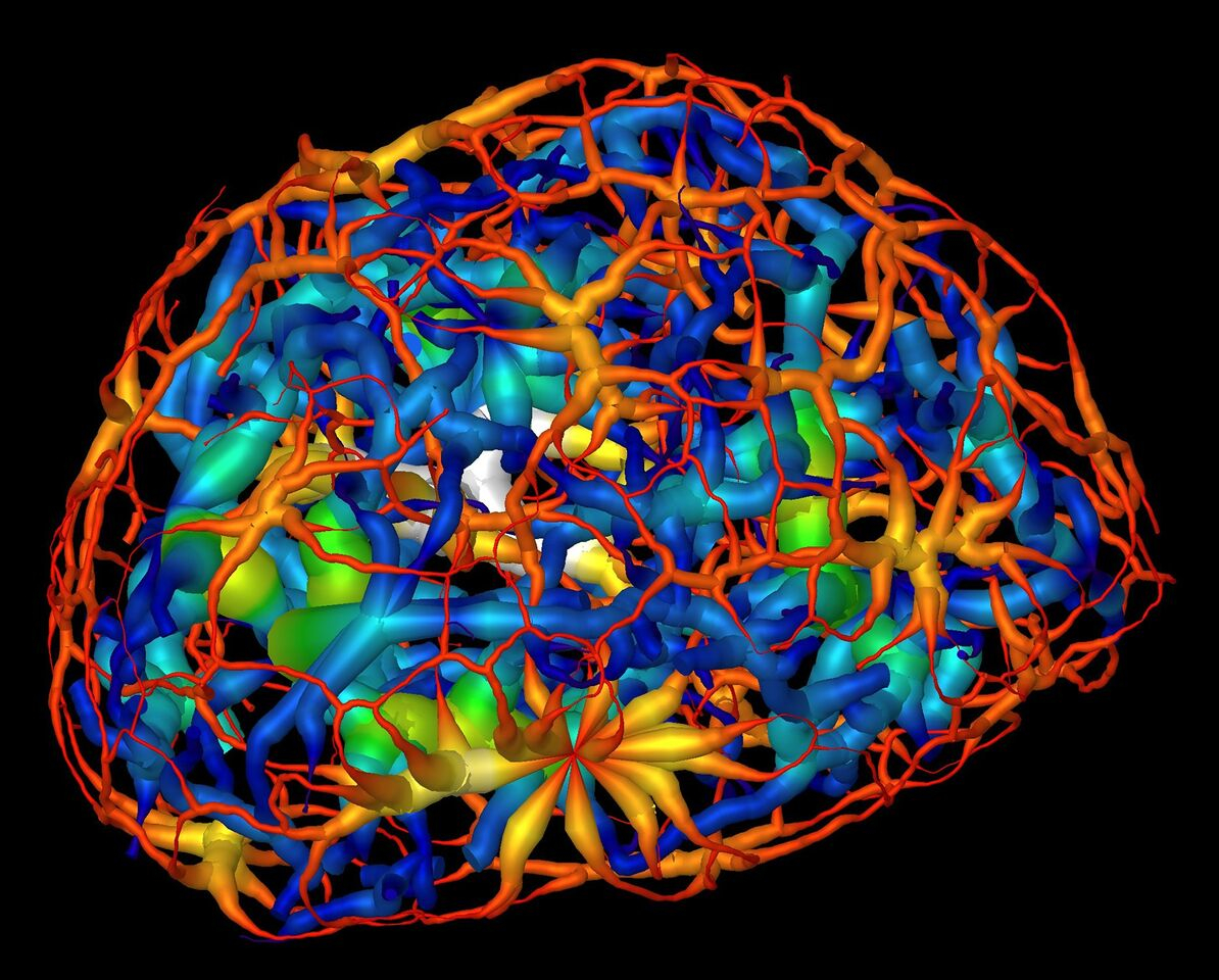
 Scientists in the Molecular Biophysics and Integrated Bioimaging Division (MBIB) have produced detailed 3-D visualizations that show an unexpected connectivity in the genetic material at the center of cells, providing a new understanding of a cell’s evolving architecture. A team of researchers, led by Carolyn Larabell, MBIB faculty scientist and professor at UCSF,
  have mapped the reorganization of genetic material that takes place when a stem cell matures into a nerve cell using X-ray imaging tools in the National Center for X-ray Tomography at the Advanced Light Source. The results could help us understand how patterning and reorganization of DNA-containing material called chromatin in a cell’s nucleus relate to a cell’s specialized function as specific genes are activated or silenced. Read more at the Berkeley Lab News Center.


