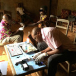
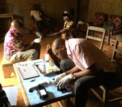
 A smartphone microscope, CellScope Loa, uses video to automatically detect and quantify infection by parasitic worms in a drop of blood. Daniel Fletcher, faculty engineer in the Biological Systems & Engineering Division, led the team of researchers who developed this next generation of UC Berkeley’s CellScope technology, which could be important for reviving efforts to eradicate debilitating filarial diseases in Africa, such as onchocerciasis (river blindness). “We previously showed that mobile phones can be used for microscopy, but this is the first device that combines the imaging technology with hardware and software automation to create a
  complete diagnostic solution,” said Daniel Fletcher, chair and professor of bioengineering at UC Berkeley (right). “The video CellScope provides accurate, fast results that enable health workers to make potentially life-saving treatment decisions in the field.” The antiparasitic drug ivermectin, or IVM, can be used to treat diseases caused by filarial worms, but mass public health campaigns to administer the medication have been stalled because of potentially fatal side effects for patients co-infected with Loa loa, which causes loiasis, or African eye worm. CellScope Loa provides critical information about high circulating levels of microscopic Loa loa worms in a patient and drastically decreases the time for diagnosis. After a successful pilot study, the team is rolling out a the new device to treat 30,000 people in Cameroon this fall. Read more at Berkeley Engineer.


