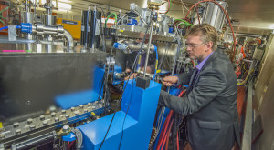
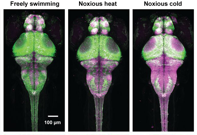
A team of scientists from the Howard Hughes Medical Institute’s Janelia Research Campus designed a fluorescent protein (CaMPARI) that causes permanent marking of active brain cells. They validated this new tool via x-ray crystallographic studies at the Berkeley Center for Structural Biology at the Advanced Light Source.  The protein was used to study live changes via fluorescence in the active nerve cells in brains of fruit flies, zebrafish, and mice. Needing to know the underlying structure of this new tool in order to understand exactly how it worked, the researchers turned to crystallography. They brought CaMPARI to Beamline 8.2.2, run by Peter Zwart (pictured) of the Molecular Biophysics and Integrated Bioimaging Division, to solve its molecular structure. Their results will help them improve CaMPARI’s sensitivity and versatility, enabling further brain activity studies. Read more at the Advanced Light Source.


