Biosciences Area

  • About Biosciences
    • Leadership
    • Area Operations Centers
    • Strategic Plan and Reports
    • Strategic Programs Development Group
    • Contact Information
  • Our Science
    • Area Programs
    • Strategic Initiatives
    • Biological Systems and Engineering
    • Environmental Genomics and Systems Biology
    • Molecular Biophysics and Integrated Bioimaging
    • DOE Joint Genome Institute
  • Media and Events
    • News
    • Announcements
    • Behind the Breakthroughs
    • Events Calendar
    • Seminar Series
  • Staff Resources
    • Commonly Used Acronyms
    • Communications
    • Hiring and Recruitment
    • Hybrid & Telework Resources
    • IDEA
    • Intellectual Property, Industry Engagement, and Entrepreneurship
    • LDRD Information
    • Logos and Templates
    • Mentoring Program
  • Search

Three Is Not a Crowd: Designed Metalloprotein Trimer Provides Stable Platform for Further Development

January 24, 2017

4iww_300
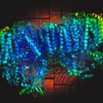
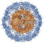
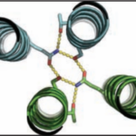
University of Washington (UW) researchers have designed a novel protein with properties that could lead to the generation of new photoactive proteins. This three-fold symmetric, self-assembling protein homotrimer contains a highly stable noncanonical amino acid. Noncanonical amino acids are not found among the 20 encoded amino acids in the body and can contain modifications to allow for new functionality. In this case, this amino acid contains a bipyridine group that chelates metal, thereby introducing new photochemical properties into the protein interface, and nucleating the formation of the homotrimer.

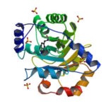
An article published last month in PNAS describes this work from a team of scientists led by David Baker at UW, which included Jose Henrique Pereira, Banumathi Sankaran, and Peter Zwart of the Molecular Biophysics & Integrated Bioimaging Division (MBIB). The MBIB scientists developed the crystal screen that was used to crystallize the novel protein and performed X-ray crystallography on Beamline 8.2.1 in the Berkeley Center for Structural Biology at the Advanced Light Source. Their X-ray crystallographic analysis of the homotrimer showed that the design process had near-atomic-level accuracy, demonstrating that computational protein design together with the utilization of noncanonical amino acids could be used to generate novel protein functions. These methods could be used to develop new therapeutics, biomaterials, and metalloproteins with useful optical or photochemical properties.

Biosciences Researchers to Support Two Exascale Projects

November 7, 2016

Data_using_LCLS X-ray laser_Sauter

Biosciences researchers will contribute their expertise to two new projects, “Data Analytics at the Exascale for Free Electron Lasers” and “Exascale Solutions for Microbiome Analysis,” funded by DOE to develop cutting-edge research applications for next-generation supercomputers as part of DOE’s Exascale Computing Project (ECP), a component of President Obama’s National Strategic Computing Initiative that intends to maximize the benefits of high-performance computing for U.S. economic competiveness, national security and scientific discovery. ECP announced its first round of funding on September 7 with the selection of 15 application development proposals for full funding and seven proposals for seed funding.

Scientists Find New Way to Manipulate Size of Virus-Like Particles

September 12, 2016

Nano Letters-virus-like particle (VLP) image

In their Nano Letters paper released August 23, 2016, a research team led by Danielle Tullman-Ercek, biologist faculty scientist in Molecular Biophysics and Integrated Bioimaging, described a new size selection method for virus-like particle assembly using chromatography. Their work has important implications for virus evolution theory, multi-protein assembly behavior, and protein-based nanomaterial development.

Bioscientists Validate Novel Protein Design Program

May 23, 2016

Over the course of billions of years, nature has evolved particular molecular structures that form the basis of life, such as those found in nucleic acids and proteins. Using the natural form as a springboard, University of Washington researchers have designed protein homo-oligomers, or identical interacting subunits, which can contain interchangeable hydrogen bonding modules for building different structures or functions. The team of researchers, led by David Baker at the University of Washington, included Jose Henrique Pereira, Banumathi Sankaran, and Peter Zwart of the Molecular Biophysics & Integrated Bioimaging Division (MBIB).

A Designed Protein Maps Brain Activity

October 28, 2015

A team of scientists from the Howard Hughes Medical Institute’s Janelia Research Campus designed a fluorescent protein (CaMPARI) that causes permanent marking of active brain cells. They validated this new tool via x-ray crystallographic studies at the Berkeley Center for Structural Biology at the Advanced Light Source.

  • « Previous Page
  • 1
  • 2
  • 3
  • 4
  • Next Page »

Was this page useful?

Send
like not like

About Biosciences

  • Leadership
  • Area Operations Centers
  • Inclusion, Diversity, Equity, and Accountability (IDEA)
  • Contact

Divisions & User Facility

  • Biological Systems and Engineering
  • Environmental Genomics and Systems Biology
  • Molecular Biophysics and Integrated Bioimaging
  • DOE Joint Genome Institute

Resources

  • A-Z Index
  • Phonebook
  • Logos
  • Acronyms
  • Integrated Safety Management
Questions & Comments
Follow us: Mastodon LinkedIn YouTube