Ke Xu
Chemist Faculty Scientist
Research Interests
Our current research focuses on the development of next-generation microscopy and spectroscopy tools to address pressing questions in cell biology. We developed a new method to enable electron microscopy, and correlated super-resolution microscopy, of fully hydrated animal cells by employing graphene, a single-atom-thick carbon meshwork, as the thinnest possible impermeable and conductive membrane to protect cells from vacuum. We also recently introduced and realized the new concept of spectrally resolved, “true-color” super-resolution microscopy, in which we synchronously obtain the fluorescence spectra and positions of millions of single molecules in densely labeled cell samples in minutes. Based on this approach, we have demonstrated in our work high-quality multicolor 3D super-resolution microscopy for 4 dyes only 10 nm separated in emission wavelength.
Recent Publications
Related News
Ke Xu Receives NIH New Innovator Award
Ke Xu, a faculty scientist in Molecular Biophysics and Integrated Bioimaging, has been awarded a NIH Director’s New Innovator Award as part of the National Institutes of Health's High-Risk, High-Reward Research program. Supported by the NIH Common Fund, these grants catalyze exceptionally innovative biomedical research with transformative potential from early career investigators who have never received an NIH grant before. An assistant professor of chemistry at UC Berkeley, Xu develops new physical and chemical tools to explore biological, chemical, and materials systems at the nanoscale with extraordinary resolution and sensitivity. He takes a multidimensional approach that integrates advanced microscopy, spectroscopy, cell biology, and nanotechnology. Xu is among 89 New Innovator Award recipients for 2018. Each grant comes with $1.5 million in direct funds for five years. Read more from UC Berkeley News.
Super-Resolution Microscopy Reveals Fine Detail of Cellular Mesh
Ke Xu, faculty scientist in the Molecular Biophysics and Integrated Bioimaging Division, used super-resolution microscopy to reveal the geodesic mesh supporting red blood cells, enabling them to be sturdy yet flexible enough to squeeze through narrow capillaries as they carry oxygen to tissues. The discovery could help uncover how malaria parasites hijack this mesh and destroy red blood cells. Read more at UC Berkeley News.
The Strings That Bind Us: Cytofilaments Connect Cell Nucleus to Extracellular Microenvironment
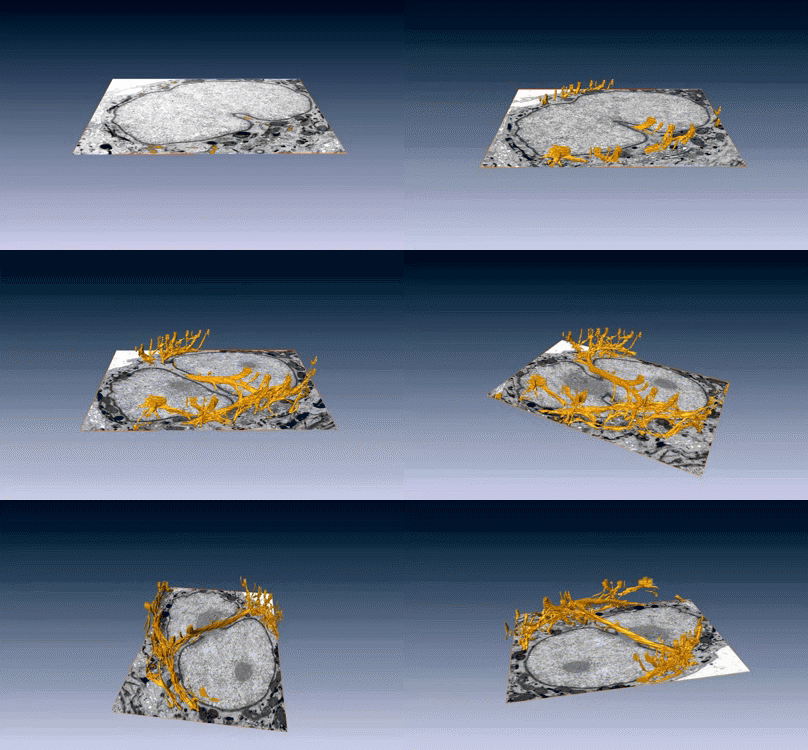
New images of structural fibers inside a cell appear in a study featured on the cover of the Journal of Cell Science special issue on 3D Cell Biology, published this month. The images, obtained by scientists in the Biosciences Area, show thread-like cytofilaments reaching into and traversing a human breast cell’s chromatin-packed nucleus.  It provides the first visual evidence of a physical link by which genes can receive mechanical cues from its microenvironment.
The work leading up to the images began in the early 1980s when Biological Systems & Engineering's Mina Bissell proposed the idea that gene expression and cell fate were dependent on their physical surroundings called extracellular matrix. The images were captured by Manfred Auer, staff scientist, and Ke Xu, faculty scientist, both in the Molecular Biophysics & Integrated Bioimaging Division. Read more at the Berkeley Lab News Center.


